Session Information
Date: Monday, November 9, 2015
Title: Imaging of Rheumatic Diseases Poster II: X-ray, MRI, PET and CT
Session Type: ACR Poster Session B
Session Time: 9:00AM-11:00AM
Background/Purpose: Inflammation of the
tendon sheaths (tenosynovitis) is a recognised component of rheumatoid
arthritis (RA). A comprehensive assessment of inflammation will require the
inclusion of tenosynovitis as well as synovitis and osteitis, and consequently
it is proposed to include a semi-quantitative assessment of wrist tenosynovitis
in the OMERACT RAMRIS scoring system.
Active appearance models (AAM) have been successfully used to develop an
automatic quantitative version of the current RAMRIS methodology.
This
study was a pilot investigation in established RA to assess whether AAMs can be
used to produce an automatic tenosynovitis measure and compare the response to
therapy of quantitative wrist tenosynovitis and synovitis measures.
Methods: MR images of the hand were
acquired at 0, 3 and 6 months from 34 established, seropositive RA patients who
received a cycle of rituximab therapy in an open label study. Pre- and post-contrast VIBE images with fat
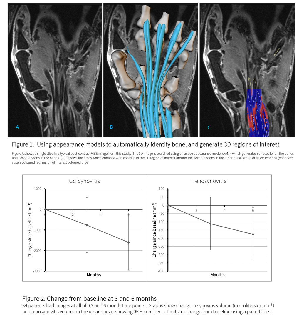
saturation were acquired, and searched with AAMs to identify bones and capsular
structures and generate 3D regions of interest (ROIs). Volume which enhanced with contrast was
calculated using a shuffle transform. AAMs of the flexor tendons were generated
from an independent training set of hand MR images. Briefly, the process includes manual
segmentation of the tendons by an expert, generating 3D surfaces using a
marching cubes algorithm and the generation of AAMs for each tendon. Images
were automatically searched using the AAM, and visually inspected to ensure
that the search process had correctly identified the tendons. A 3D region of interest (ROI) around each
tendon was created by inflating the tendon shape to form a halo which included
the tendon sheath. Within the ROI the
tenosynovitis volume was calculated using the shuffle transform method. For
this pilot study only the wrist flexor tendons within the common synovial
sheath were analysed. The amount of
change for the 2 methods was judged using a paired t-test.
Results:
Tenosynovitis
in the flexor tendons, and synovitis volume decreased at 3 and 6 months, in an
approximately linear fashion. Change was
significant at 6 months for both measures (Figure 2). Although the change in
the population mean was linear for both measures, the slope of change in tenosynovitis volume
for individual patients did not correlate with change in synovitis volume for
the same patients (r2 = 0.29).
Conclusion:
It
is feasible to quantify tenosynovitis using AAMs. Tenosynovitis in the flexor tendons decreased
over 6 months, and only correlated weakly with change in synovitis volume
within the same patient. Tenosynovitis appears to be as responsive as synovial volume,
but did not correlate with synovial change in individual patients in this small
study. Tenosynovitis may therefore add
new information to that already provided by measures of synovitis, though this
will need confirmation with a fully developed tool in a larger RA population.
To cite this abstract in AMA style:
Bowes MA, Guillard G, Vincent GR, Freeston JE, Vital EM, Emery P, Conaghan PG. Quantitative MRI Measurement of Tenosynovitis Demonstrates Differing Responses of Synovitis and Tenosynovitis after RA Treatment [abstract]. Arthritis Rheumatol. 2015; 67 (suppl 10). https://acrabstracts.org/abstract/quantitative-mri-measurement-of-tenosynovitis-demonstrates-differing-responses-of-synovitis-and-tenosynovitis-after-ra-treatment/. Accessed .« Back to 2015 ACR/ARHP Annual Meeting
ACR Meeting Abstracts - https://acrabstracts.org/abstract/quantitative-mri-measurement-of-tenosynovitis-demonstrates-differing-responses-of-synovitis-and-tenosynovitis-after-ra-treatment/

