Session Information
Date: Sunday, November 8, 2015
Title: Systemic Lupus Erythematosus - Clinical Aspects and Treatment Poster Session I
Session Type: ACR Poster Session A
Session Time: 9:00AM-11:00AM
Background/Purpose:
Systemic lupus erythematosus (SLE) is a prototypic autoimmune disease characterized by immune complexes of antinuclear antibodies. As a source of these antigens, increased levels of apoptosis and defective clearance of dead cells have been proposed. As now recognized, during apoptosis and cell activation, microvesicles (MVs) are released extra-cellularly. MVs are small membrane bound vesicles that enclose intracellular contents. To determine whether MVs contain nuclear molecules and can form immune complexes, we quantified by flow cytometry the content of nucleic acids; High Mobility Group Box-1 (HMGB1), a non-histone protein with alarmin activity; and bound immunoglobulin on MVs from SLE patients and controls.
Methods:
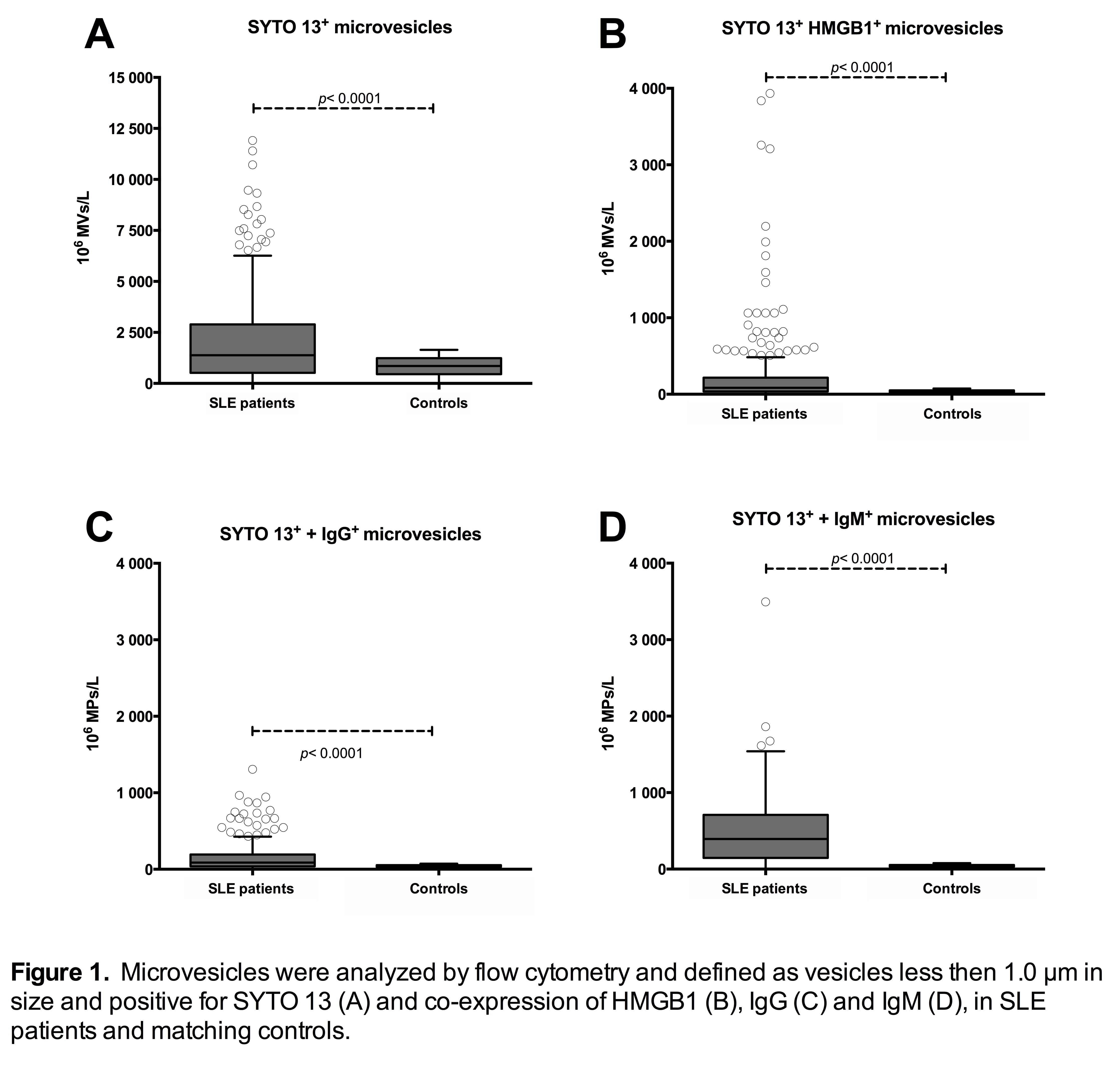
Fasting plasma samples from 294 patients with SLE (4≥ of 1982 ACR criteria) and 309 age- and sex-matched population controls were investigated. MVs were analyzed by flow cytometry as vesicles less than 1.0 µm in size and positive for SYTO 13, a cell permeable dye binding to DNA or RNA. We also measured expression of HMGB1, IgG and IgM on MVs.
To determine whether factors in SLE blood can bind MVs, MVs from healthy volunteers were incubated for 60 min at room temperature with plasma from SLE patients (n=5). After incubation, the MVs were isolated and the amounts of IgG and IgM were measured.
Results:
Blood of SLE patients had significantly higher numbers of MVs containing nucleic acids and these MVs expressed more HMGB1, IgG and IgM as compared to controls (Figure 1A-D).
In-vitro experiments revealed that incubation of MVs obtained from healthy volunteers with MV-free plasma obtained from SLE patients, significantly increased the levels of IgG and IgM on MVs (IgG: 0.3±0.03 vs 0.64±0.07 mean fluorescence intensity (MFI); IgM: 0.29±0.02 vs 0.41±0.08 MFI; p<0.0001 and p<0.05 respectively).
Conclusion:
MVs containing nucleic acids are significantly more abundant in the blood of SLE patients than in controls. Many of these MVs expose the alarmin HMGB1, IgG and in particular, IgM on their surface. Furthermore, plasma from SLE patients increased immunoglobulin binding on MVs from controls. Together, these studies indicate that release of MVs into the blood is a feature of SLE and that immunoglobulins can bind to antigenic sites on MVs. Since MVs are plentiful, form immune complexes, and display an alarmin, these vesicles could drive SLE pathogenesis as a source of immune complexes and immunostimulatory molecules such as HMGB1.
To cite this abstract in AMA style:
Mobarrez F, Wallén H, Gunnarsson I, Gustafsson J, Zickert A, Pisetsky D, Svenungsson E. Microvesicles Containing Nucleic Acids and Expressing Immunoglobulins and HMGB1 Are Abundant in Patients with Systemic Lupus Erythematosus [abstract]. Arthritis Rheumatol. 2015; 67 (suppl 10). https://acrabstracts.org/abstract/microvesicles-containing-nucleic-acids-and-expressing-immunoglobulins-and-hmgb1-are-abundant-in-patients-with-systemic-lupus-erythematosus/. Accessed .« Back to 2015 ACR/ARHP Annual Meeting
ACR Meeting Abstracts - https://acrabstracts.org/abstract/microvesicles-containing-nucleic-acids-and-expressing-immunoglobulins-and-hmgb1-are-abundant-in-patients-with-systemic-lupus-erythematosus/

