Session Information
Date: Sunday, November 8, 2015
Title: Osteoarthritis - Clinical Aspects Poster I: Treatments and Metabolic Risk Factors
Session Type: ACR Poster Session A
Session Time: 9:00AM-11:00AM
Background/Purpose: Knee
osteoarthritis (OA) is a degenerative disease associated with significant
muscle weakness and disability. Ectopic fat in the thigh, including
intramuscular fat (intraMF; fat within a muscle belly) and intermuscular fat
(IMF; fat within the deep fascia and between muscles bellies), is associated
with impaired strength and physical performance in knee OA. Despite the role of
calf muscles in creating power for gait and mobility, there has been little investigation
of calf muscle composition in knee OA. We investigated the relationships
between calf muscle adiposity, IMF and intraMF volume with physical performance
in women with knee OA.
Methods:
Women
(n=20) with radiographic knee OA had the calf of their most symptomatic knee
imaged using 3.0T magnetic resonance imaging (MRI). The iterative decomposition
of water and fat with echo asymmetry and least-squares estimation (IDEAL)
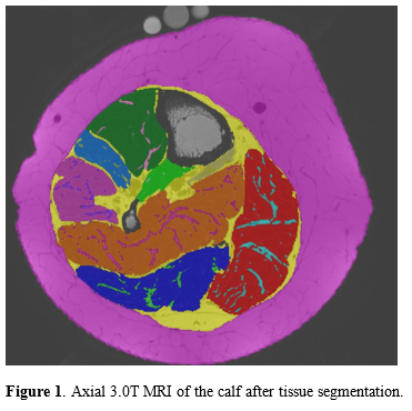
sequence obtained 10 fat-separated axial images (3 mm slice thickness). Images
were analyzed using SliceOmatic® software with region-growing to quantify IMF,
intraMF and lean muscle volumes (cm3) (Figure 1). Muscle adiposity
was calculated as the percent of total muscle volume consisting of intramuscular
fat; this was calculated for the whole calf and individually for the medial
gastrocnemius (MG), lateral gastrocnemius (LG), soleus (SOL), and tibialis
anterior (TA). Participants performed five physical performance tests, and rated
their knee pain using a numeric pain rating scale. Linear regression analyses
were performed.
Results:
Mean
sample characteristics (±SD): age 65±5 y; BMI 30±5 kg/m2. After
adjusting for knee pain, whole calf muscle adiposity, but not IMF or intraMF volume,
was associated with poor performance on the timed up-and-go (TUG), stair
ascent/descent test (SADT), and 6 minute walk test (6WT) (Table 1).
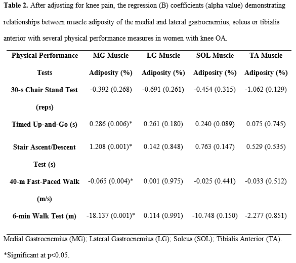
Furthermore, MG adiposity, but not LG, SOL or TA adiposity, was associated with
poor performance on the TUG, SADT, 6WT, and 40 meter fast-paced walk test
(40FPWT) (Table 2).
Conclusion:
Whole
calf, and particularly medial gastrocnemius adiposity, are associated with poor
physical performance in women with knee OA. The medial gastrocnemius may be
important in mobility disability in OA. Further investigation of longitudinal
changes in calf muscle composition in knee OA are required.
To cite this abstract in AMA style:
Davison M, Maly MR, Adachi JD, Beattie KA. Calf Muscle Adiposity Is Associated with Impaired Physical Performance in Knee OA [abstract]. Arthritis Rheumatol. 2015; 67 (suppl 10). https://acrabstracts.org/abstract/calf-muscle-adiposity-is-associated-with-impaired-physical-performance-in-knee-oa/. Accessed .« Back to 2015 ACR/ARHP Annual Meeting
ACR Meeting Abstracts - https://acrabstracts.org/abstract/calf-muscle-adiposity-is-associated-with-impaired-physical-performance-in-knee-oa/


