Session Information
Session Type: Poster Session D
Session Time: 1:00PM-3:00PM
Background/Purpose: YKL-40 is a chitinase-like protein that is associated with interstitial lung disease in patients with polymyositis (PM)/dermatomyositis (DM) but not with myositis. Myositis pathogenesis is complex, and atypical myositis is clinically classified as PM/DM albeit with reduced muscle weakness. Here, we investigated whether YKL-40 is associated with PM/DM and assessed its role in PM/DM pathogenesis.
Methods: Thirty-five patients diagnosed with PM/DM based on Bohn and Peter’s classification criteria were retrospectively enrolled and evaluated at Hyogo Medical University Hospital from 2006 to 2020 and the results were compared with those of 26 healthy controls (HC). Patients satisfied the 2017 EULAR/ACR classification criteria for idiopathic inflammatory myopathies (IIM). Serum YKL-40 levels were measured by enzyme-linked immunosorbent assay (ELISA) and age-corrected to YKL-40 percentile values. The correlation between age corrected YKL-40 percentile and serum markers [creatine kinase (CK), C-reactive protein (CRP), and lactic dehydrogenase (LDH)] was determined using the Mann-Whitney U test. Muscle biopsies obtained from two PM and two DM patients were subjected to immunohistochemistry and immunofluorescence staining using anti-YKL-40 and anti-CD68 antibodies to identify inflammatory cells expressing YKL-40.
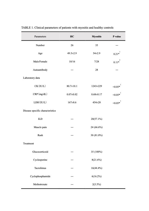
Results: The mean age, sex ratio, number of patients, symptoms, clinical data, and treatment are shown in TABLE 1. Age-corrected serum YKL-40 values were significantly increased in patients with PM/DM compared to the HC but significantly decreased before and after treatment. Serum YKL-40 levels weakly correlated with CRP but not with CK or LDH. Immunohistochemical analysis showed infiltration of YKL-40-positive inflammatory cells in the endomysium and perimysium of the muscle sheath. Immunofluorescence staining revealed CD68 expression in YKL-40-positive inflammatory cells, suggesting that these cells were macrophages.
Conclusion: We believe that the trend of serum YKL-40 levels in PM/DM patients can reflect myositis. Serum CK and LDH levels indicate muscle destruction as they are derived from muscles. Serum YKL-40 levels did not correlate with serum CK and LDH levels, possibly due to the inclusion of cases with mild muscle destruction. Thus, YKL-40 may be used to evaluate myositis cases with mild muscle damage. CD8+ and CD4+ T cells are believed to play predominant roles in muscle destruction in PM and DM, respectively. However, YKL-40-positive macrophages observed in both diseases suggest that cells other than CD8+ and CD4+ T cells may cause inflammation. This provides new insights to understand pathophysiological mechanisms of atypical myositis.
To cite this abstract in AMA style:
Noguchi K, Furukawa T, Yoshikawa T, Morimoto M, Hashimoto T, Azuma N, Matsui K. YKL-40-mediated Inflammatory Pathogenesis Common to Polymyositis / Dermatomyositis [abstract]. Arthritis Rheumatol. 2022; 74 (suppl 9). https://acrabstracts.org/abstract/ykl-40-mediated-inflammatory-pathogenesis-common-to-polymyositis-dermatomyositis/. Accessed .« Back to ACR Convergence 2022
ACR Meeting Abstracts - https://acrabstracts.org/abstract/ykl-40-mediated-inflammatory-pathogenesis-common-to-polymyositis-dermatomyositis/

