Session Information
Date: Sunday, November 12, 2023
Title: (0229–0251) Metabolic & Crystal Arthropathies – Basic & Clinical Science Poster I
Session Type: Poster Session A
Session Time: 9:00AM-11:00AM
Background/Purpose: The gold standard for diagnosis of crystal arthritis relies on the identification of either monosodium urate (MSU) or calcium pyrophosphate (CPP) crystals in synovial fluid by compensated polarized light microscope (CPLM). However, CPLM analysis is labor intensive and depends on experience of the technician. Identifying CPP crystals can be difficult. We previously described single-shot computational polarized light microscopy (SCPLM), that detects MSU and CPP crystals in synovial fluid. This work evaluates the reliability and validity of crystal detection for SCPLM images using crystal experts.
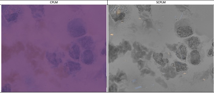
Methods: Microscope slides from patients with CPP or MSU crystals in synovial fluid were obtained and de-identified from the hospital clinical lab. Digital images were acquired using Olympus IX83 microscope, standard objective lens (100x/1.4NA) and either CPLM or SCPLM methodology. Briefly, SCPLM uses a CMOS sensor where each pixel is integrated with a directional polarizing filter with four axes of polarization (0°, 90°, 45°, 135°). SCPLM further combines images from multiple focal depths into a single bright-field fused image.
In random order, raters were presented paired CPLM images (with analyzer at orthogonal angles) and a single bright-field fused SCPLM image for 67 FOV (including 7 negative controls). For each FOV and each method, each rater recorded their level of certainty (1-5) for each suspect crystal identified and type of crystal. Crystals rated 3 or higher by both raters (++) on either method were included in a high-certainty crystal subset. After rating all of the FOV, raters were presented with side-by-side FOV (CPLM vs. SCPLM) and asked for their preferred image.
Results: 67 FOV were imaged by CPLM and SCPLM methodologies (29 CPP, 31 MSU and 7 negative controls). With an instructed limit of 15 crystals per FOV, 377 unique crystal suspects were identified: 280 CPP, 87 MSU, and 10 uncertain or discrepant crystal identity. (The 10 uncertain/discrepant crystals all came from negative control FOVs.)
Raters identified a higher number of crystals by SCPLM over CPLM for both CPP and MSU. (Table 1) SCPLM identified 239/280 CPP crystals and 86/87 MSU crystals where CPLM identified 138/280 CPP and 48/87 MSU. For SCPLM, there were only 11/239 CPP crystals (4.6%) where neither rater was certain, for CPLM, there were 13/138 (9.4%) where neither rater was certain.
To compare methods, we focused on the 144 CPP and 69 MSU crystals where both raters were certain (++) about the crystal (by at least one method). For 80-90% of included crystals, SCPLM was ++. In contrast, only 40-51% of crystals was CPLM ++. (Table 2) When CPLM was negative, SCPLM was high certain in almost all cases.
Finally, raters subjectively preferred SPCLM over CPLM in side-by-side comparison. Raters were indifferent to method for negative FOVs.
Conclusion: Subjective (rater preference) and objective measures of greater detection and higher certainty were observed for SCPLM images over standard CPLM images, particularly notable for CPP crystals. The digital data associated with these images can be incorporate into an automated scanning platform to provide quantitative reports on crystal counts and morphology perhaps providing greater insight to clinical care.

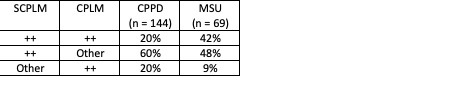
++ = both raters with high certainty about crystal for a specific method
Legend: CPLM = Compensated Polarized Light Microscopy. SCPLM = Single-shot Computational Polarized Light Microscopy
To cite this abstract in AMA style:
FitzGerald J, Liu T, Barrios C, Bai B, McCarthy G, Rosenthal A, Ozcan A. Validation of Singe-Shot Computational Polarized Microscopy for Crystal Analysis of Synovial Fluid [abstract]. Arthritis Rheumatol. 2023; 75 (suppl 9). https://acrabstracts.org/abstract/validation-of-singe-shot-computational-polarized-microscopy-for-crystal-analysis-of-synovial-fluid/. Accessed .« Back to ACR Convergence 2023
ACR Meeting Abstracts - https://acrabstracts.org/abstract/validation-of-singe-shot-computational-polarized-microscopy-for-crystal-analysis-of-synovial-fluid/