Session Information
Session Type: Poster Session C
Session Time: 9:00AM-11:00AM
Background/Purpose: Giant cell arteritis (GCA) is the most prevalent type of primary systemic vasculitis among individuals aged 50 and above. Imaging techniques such as ultrasound (US), magnetic resonance imaging (MRI), and {18F}-fluorodeoxyglucose positron emission tomography (PET-CT) have emerged as crucial diagnostic tools due to their non-invasive nature, higher sensitivity and availability compared to temporal artery biopsy (TAB) and angiography.
The purpose of this study is to evaluate the effectiveness of an algorithm based on imaging techniques in the diagnosis of GCA. Additionally, it seeks to assess the potential reduction in costs and unnecessary investigations during the diagnostic process.
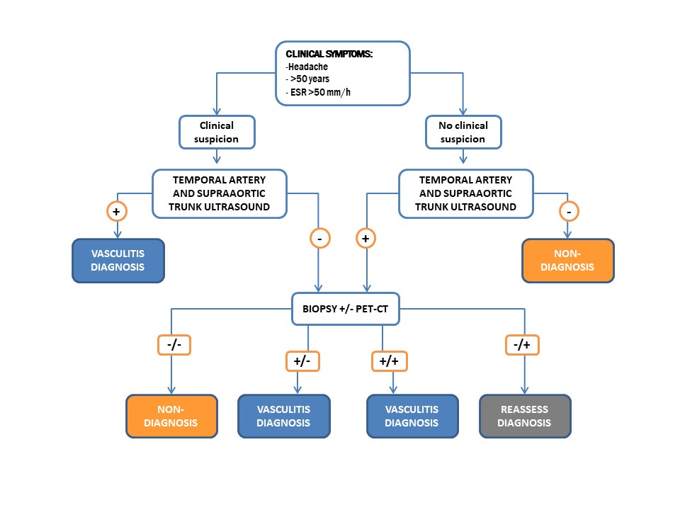
Methods: The project consists of two phases. The first phase involved a retrospective analysis of a cohort suspected of having GCA, derived from outpatient consultations (OC) and hospitalizations in a tertiary hospital’s Rheumatology department (February 2021-December 2022). A diagnostic evaluation was conducted using clinical criteria and imaging techniques (US, PET-CT, TAB). US examinations were performed according to a standardized protocol within the Rheumatology service. In the second phase, the proposed diagnostic algorithm (Figure) was retrospectively applied, and the results were compared with the pre-algorithm phase. All patients provided their consent for participation.
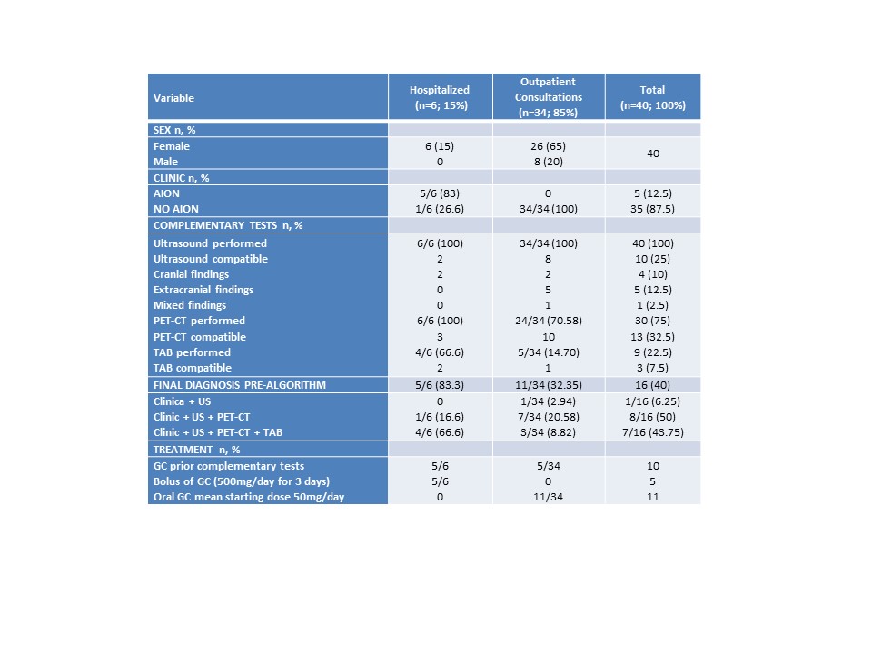
Results: The study included 40 patients (80% women, n=32; 20% men, n=8), with a mean age of 72.55. All patients had clinical suspicion and 100% underwent US examinations with findings suggestive of GCA in 10 individuals (25%). TAB was performed in 9 patients (22.5%) and 3 of them (7.5%) had compatible results. PET-CT was conducted in 30 (75%) with conclusive results observed in 13 cases (32.5%). A final diagnosis of GCA was established in 16 patients (40%): clinical criteria + US in 1 patient (6.25%); clinical criteria + US + PET-CT in 8 (50%); clinical criteria + US + PET-CT + TAB in 7 (43.75%) (Table). Notably, among the 6 patients admitted to the hospital, 5 presented with initial clinical manifestations of anterior ischemic optic neuritis (AION). Within the diagnosed group, 10 individuals received corticosteroid treatment for at least 5 days prior to undergoing imaging techniques. Retrospective application of the algorithm to 8 patients (20%) with compatible US findings indicated that no further investigations would have been necessary (TAB + PET-CT in 5 patients and PET-CT alone in 3). Consequently, a total of 13 unnecessary supplementary tests could have been avoided. The maximum cost per patient amounted to €1117 (PET-CT €997, US €70, TAB €50). Implementation of the algorithm could have resulted in cost savings of €8176 and reduced iatrogenesis in this subgroup.
Conclusion: The utilization of an algorithm represents a practical and applicable tool in the diagnostic process of GCA. US serves as a valuable screening method in the initial evaluation, effectively reducing costs and redundant investigations in patients with compatible results and a high clinical suspicion. In cases where both clinical criteria and US findings are negative, it aids in excluding GCA as a diagnosis.
To cite this abstract in AMA style:
AVALOS BOGADO H, AÑEZ STURCHIO G, Trallero-Araguas E, Espartal López E, Sandoval Moreno s, Ulloa Navas d, De Agustín De Oro J. Utility of a Diagnostic Algorithm in the Assessment of Large Vessel Vasculitis [abstract]. Arthritis Rheumatol. 2023; 75 (suppl 9). https://acrabstracts.org/abstract/utility-of-a-diagnostic-algorithm-in-the-assessment-of-large-vessel-vasculitis/. Accessed .« Back to ACR Convergence 2023
ACR Meeting Abstracts - https://acrabstracts.org/abstract/utility-of-a-diagnostic-algorithm-in-the-assessment-of-large-vessel-vasculitis/


