Session Information
Title: Systemic Lupus Erythematosus - Clinical Aspects and Treatment: Treatment and Management Studies
Session Type: Abstract Submissions (ACR)
Background/Purpose: Ionizing radiation from CT scanning can increase cancer risk. Lupus patients are frequently evaluated for chest pain and may have multiple pulmonary embolism CT (PE-CT) scans in addition to other exposures to diagnostic or therapeutic radiation.
The objective of this study is twofold i) Determine the incidence of PE in University of Michigan Lupus Cohort patients who have undergone PE -CT scans ii) Estimate associated breast and lung cancer incidence
Methods: We reviewed records of patients in the Michigan Lupus Cohort (N=856), and for each patient determined the number and outcome of PE-CT scans. All patients gave informed consent for review of their records. Based on estimated x-ray exposure from a state of the art GE Discovery CT750 HD CT scanner for an arterial chest scan, we estimated radiation dosage to the breast and lung in an average sized adult woman, utilizing CT-Expo software package.{1] We used the dose information and the patient’s age at the time of the CT scan and tabulated risks according to the BEIR VII report to estimate the increased lifetime incidence risks of breast and lung cancer. Risks from multiple CT scans were summed.
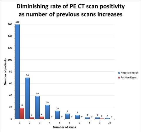
Results: 183/856 (21%) patients underwent a total of 358 PE CT scans. The overall rate of positivity was 7.5%. The likelihood of a positive scan decreased in proportion to the number of scans that had been previously performed. (Figure 1) For patients undergoing their first through third CT scans the rate of positivity for PE was 9.2 %, whereas patients undergoing their fourth through tenth CT scans had 1.6% positivity.
We estimated radiation doses of to the breast and lung in female patients from a PE CT arterial scan, as 20mGy and 22mGy respectively. Separating patients based on age and number of CT scans, the estimated lifetime increase incidence risk of breast and lung cancer was calculated. In this simplified model the range of added risk for breast and lung cancer respectively per 100,000 female patients was 0-100 in 72% and 72%; 100- 500 in 25% and 28% and 500- 1000 in 4% and 1%.
Image/graph:
Conclusion: Patients with multiple previous PE CT scans had lower likelihood of a positive result on subsequent scans and higher risks of malignancy. Our conservative estimates of radiation exposure, that do not account for higher radiation doses from older CT scanners, indicate sufficient risk to warrant more precise characterization of radiation exposure in this population; and encourage development of guidelines for PE screening for patients with systemic lupus erythematosus and recurrent chest pain. The magnitude of risk should not discourage performance of PE CT when clinically indicated.
References: G. Stamm and H. D. Nagel, “CT-expo—A novel program for dose evaluation in CT,” RoFo: Fortschritte auf dem Gebiete der Ro¨ntgenstrahlen und der Nuklearmedizin 174, 1570–1576 (2002)
Disclosure:
R. Kado,
None;
E. Siegwald,
None;
E. Lewis,
None;
M. Goodsitt,
None;
E. Christodoulou,
None;
E. Kazerooni,
None;
W. J. McCune,
None.
« Back to 2014 ACR/ARHP Annual Meeting
ACR Meeting Abstracts - https://acrabstracts.org/abstract/utility-and-associated-risk-of-pulmonary-embolism-ct-scans-in-the-michigan-lupus-cohort/

