Session Information
Date: Sunday, October 26, 2025
Title: (0233–0279) Miscellaneous Rheumatic & Inflammatory Diseases Poster I
Session Type: Poster Session A
Session Time: 10:30AM-12:30PM
Background/Purpose: Small fiber neuropathy (SFN) is a common complication of sarcoidosis, affecting about one third of patients. The current gold standard for diagnosis combines patient history and a skin biopsy to quantify small nerve fibers. Corneal confocal microscopy (CCM) is now being used with more frequency to evaluate nerve fibers in the cornea, specifically in diabetic neuropathy. However, further research is needed to validate the role of CCM in sarcoidosis. Our study focused on CCM and how it can be utilized as an accurate, non-invasive diagnostic tool for SFN in patients with sarcoidosis.
Methods: This study is a subset analysis of a larger study to better characterize SFN in sarcoidosis. A total of 12 patients with sarcoidosis who completed CCM analysis were included in the study. We categorized sarcoidosis patients into two groups: those with a physician impression of neuropathy (PIN) and those without a physician impression of neuropathy (non-PIN). A third group, healthy age-matched controls without sarcoidosis from a database, were added for comparison. CCM was performed to determine corneal nerve fiber density, length and tortuosity and corneal nerve branch density.
Results: CCM data was collected on 12 patients with sarcoidosis, 8 of who had PIN and 4 of who had non-PIN. The mean age of patients in the PIN group was 52.8 years with 87.5% females. The non-PIN group averaged 47.25 years old with 50% females. The healthy control group had a mean age of 51.3 years with no data on sex. One-way ANOVA analysis revealed a difference in corneal nerve fiber density (CNFD) approaching significance (p = 0.05) between the three groups. Post-hoc comparison using two-sample t-test to assess for between-group differences revealed a statistically significant difference in CNFD (p = 0.03), and a difference in corneal nerve branch density approaching statistical significance (p = 0.05) between age-matched controls and PIN patients. However, there was not a difference in corneal nerve fiber length or corneal nerve fiber tortuosity between the two groups. There was no statistical difference noted in any of the measures between healthy controls and the non-PIN group or when comparing the PIN and the non-PIN groups.
Conclusion: CCM is a non-invasive method to capture images of small nerve fibers as a marker for SFN. Primarily used in diabetic neuropathy, our study findings indicate CCM can provide diagnostic data in patients with sarcoidosis and features of SFN, especially when analyzing CNFD. Interestingly, though not statistically significant, non-PIN patients trended toward having low corneal nerve fiber and branch densities, similar to PIN patients. Due to the low number of patients in our study, further research will be needed to validate the use of CCM as a screening tool for SFN in sarcoidosis patients.
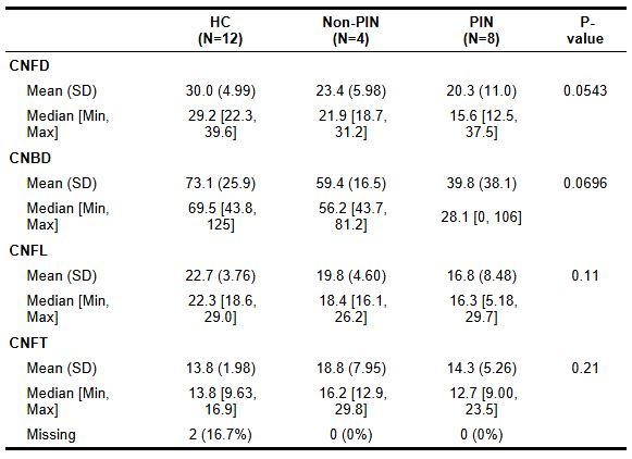 Table 1. One-way ANOVA test analyzing group differences in corneal nerve fiber density (CNFD), corneal nerve branch density (CNBD), corneal nerve fiber length (CNFL), and corneal nerve fiber tortuosity (CNFT) among three groups: health controls (HC), patients without a clinical impression of peripheral neuropathy (non-PIN), and patients with a clinical impression of neuropathy (PIN). The difference in CNFD is approaching statistical significance between the groups (p = 0.05).
.jpg) Figure 1. Corneal nerve fiber densities (CNFD) among healthy controls (HC), patients without a clinical impression of peripheral neuropathy (non-PIN), and patients with a clinical impression of neuropathy (PIN). A statistical difference was noted between healthy controls and PIN groups (p= 0.03).
Figure 1. Corneal nerve fiber densities (CNFD) among healthy controls (HC), patients without a clinical impression of peripheral neuropathy (non-PIN), and patients with a clinical impression of neuropathy (PIN). A statistical difference was noted between healthy controls and PIN groups (p= 0.03).
.jpg) Figure 2. Corneal nerve branch densities (CNBD) among healthy controls (HC), patients without a clinical impression of peripheral neuropathy (non-PIN), and patients with a clinical impression of neuropathy (PIN). Though no statistically significant difference was found among the groups, there was a trend towards significance between HC and PIN groups (p = 0.05).
Figure 2. Corneal nerve branch densities (CNBD) among healthy controls (HC), patients without a clinical impression of peripheral neuropathy (non-PIN), and patients with a clinical impression of neuropathy (PIN). Though no statistically significant difference was found among the groups, there was a trend towards significance between HC and PIN groups (p = 0.05).
To cite this abstract in AMA style:
Bucur P, Gwathmey K, Syed H, Syed A, Iden T, Gupta N, McLaughlin J, Kron J, Canissario R, Chen S, Gad H, Malik R, Patel S, Patel V. Use of Corneal Confocal Microscopy to Assess Small Fiber Neuropathy in Patients with Sarcoidosis [abstract]. Arthritis Rheumatol. 2025; 77 (suppl 9). https://acrabstracts.org/abstract/use-of-corneal-confocal-microscopy-to-assess-small-fiber-neuropathy-in-patients-with-sarcoidosis/. Accessed .« Back to ACR Convergence 2025
ACR Meeting Abstracts - https://acrabstracts.org/abstract/use-of-corneal-confocal-microscopy-to-assess-small-fiber-neuropathy-in-patients-with-sarcoidosis/