Session Information
Session Type: Abstract Submissions (ACR)
Background/Purpose: The diagnosis of calcium pyrophosphate crystal (CPP) deposition disease (CPPD) is mainly based on the synovial fluid analysis and Xrays. US has demonstrated high sensitivity and specificity values for diagnosing CPPD compared to synovial fluid analysis as the gold standard, but less is known about sensitivity and specificity of synovial fluid analysis itself. Aim of the study is to compare ultrasonography, synovial fluid analysis and X-rays performances in the diagnosis of CPPD using a real gold standard.
Methods: We enrolled in our study all patients waiting to undergo knee replacement surgery due to severe osteoarthritis. Each patient underwent US examination of the knee, focusing on the menisci and the hyaline cartilage, the day prior to surgery, scoring each site according to the presence/absence of CPP as defined previously. The day of the surgery, synovial fluid of the knee (if present) was aspirated by the surgeon. After surgery, the menisci, condyles and the synovial fluid were retrieved and examined microscopically. Synovial fluid analysis was performed on wet preparations. For the meniscus and cartilage microscopic analysis, six samples were collected, either from the surface and from the internal of the structure trying to cover a large part of it. All slides were observed under transmitted light microscopy and by compensated polarised microscopy. A dichotomous score was given for the presence/absence of CPP. US and microscopic analysis were performed by different operators, blind to each other’s findings. X-rays of the knees were collected and assessed for the presence of CPPD by a Radiologist expert in musculo-skeletal imaging, blind to other findings. Sensitivity and specificity of US, synovial fluid and X-Rays were calculated using microscopic findings of the menisci and cartilage as the gold standard.
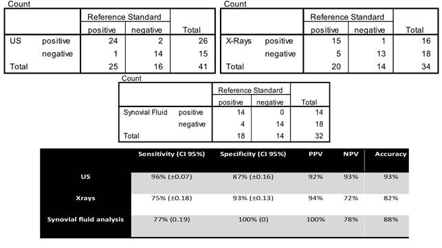
Results: we enrolled in the study 42 patients (14 males), mean age of 74 years old (±8.4). All patients underwent US of the knee, synovial fluid was present in 32 patients and X-rays have been collected form 34 patients. 2×2 contingency tables and diagnostic accuracy values for each exam are summarized in table 1.
Conclusion: US demonstrated higher sensitivity values for identifying CPP deposits in the knee joint than synovial fluid analysis. Specificity values on the other hand were higher for the microscopic analysis as expected. Globally we believe that for its intrinsic characteristics, the non invasive nature, for the high values of both specificity and specificity, and last but not least, for the capability to address differential diagnosis US should be the first exam to be performed when CPPD disease is suspected. As this study demonstrates, the presence of CPP crystals in the synovial fluid, definitely confirms the diagnosis but a negative microscopic exam does not exclude it.
Disclosure of Interest: None Declared
Disclosure:
G. Filippou,
None;
A. Adinolfi,
None;
S. Lorenzini,
None;
I. Bertoldi,
None;
V. Di Sabatino,
None;
V. Picerno,
None;
L. Sconfienza,
None;
M. Galeazzi,
None;
B. Frediani,
None.
« Back to 2014 ACR/ARHP Annual Meeting
ACR Meeting Abstracts - https://acrabstracts.org/abstract/ultrasound-versus-x-rays-versus-synovial-fluid-analysis-for-the-diagnosis-of-calcium-pyrophosphate-dihydrate-deposition-disease-is-it-cppd/

