ACR Meeting Abstracts

ACR Meeting Abstracts

  • Meetings
    • ACR Convergence 2024
    • ACR Convergence 2023
    • 2023 ACR/ARP PRSYM
    • ACR Convergence 2022
    • ACR Convergence 2021
    • ACR Convergence 2020
    • 2020 ACR/ARP PRSYM
    • 2019 ACR/ARP Annual Meeting
    • 2018-2009 Meetings
    • Download Abstracts
  • Keyword Index
  • Advanced Search
  • Your Favorites
    • Favorites
    • Login
    • View and print all favorites
    • Clear all your favorites
  • ACR Meetings

Abstract Number: 2229

Ultrasound Localization Microscopy as a New Tool for Takayasu’s Arteritis Activity Assessment, a Deep Insight Within Carotid Wall Inflammation

Guillaume Goudot1, Anatole Jimenez2, Nassim Mohamedi3, jonas Sitruk3, Lina Khider1, Hélène Mortelette3, Clément Papadacci2, Fabien Hyafil4, Mickaël Tanter2, Emmanuel Messas1, Mathieu Pernot2 and Tristan Mirault1, 1Vascular medicine department, Georges Pompidou European hospital, APHP, Université Paris Cité, INSERM U970 PARCC, Paris, Ile-de-France, France, 2Physics for Medicine Paris, INSERM U1273, ESPCI Paris, CNRS FRE 2031, PSL Research University, Paris, France, 3Vascular medicine department, Georges Pompidou European hospital, APHP, Université Paris Cité, Paris, Ile-de-France, France, 4Nuclear medicine department, Georges Pompidou European hospital, APHP, Universite Paris Cite, INSERM U970 PARCC, Paris, Ile-de-France, France

Meeting: ACR Convergence 2022

Keywords: Biomarkers, Disease Activity, Takayasu.s arteritis, Ultrasound, Vasculitis

  • Tweet
  • Click to email a link to a friend (Opens in new window) Email
  • Click to print (Opens in new window) Print
Session Information

Date: Monday, November 14, 2022

Title: Abstracts: Vasculitis – Non-ANCA-Associated and Related Disorders II

Session Type: Abstract Session

Session Time: 3:00PM-4:30PM

Background/Purpose: Use of sulphur hexafluoride microbubbles (MB) with ultrasound allow to assess arterial wall vascularization through signal enhancement. The use of ultrafast imaging with MB super-localization provides access to ultrasound localization microscopy (ULM) and thus to visualize carotid vasa vasorum in vivo. We aim to perform vasa vasorum imaging within the carotid wall in Takayasu’s arteritis and to provide a correlation with disease’s activity.

Methods: Patients with Takayasu’s arteritis were consecutively included in the French national referral centre for rare vascular diseases. Assessment of activity was performed using NIH activity index, with measurement of biological inflammation, morphological assessment by computed tomography (CT) angiography, and 18-fluorodeoxyglucose CT scan. Ultrafast ultrasound imaging was performed with a 7 MHz central frequency linear probe. A dedicated contrast imaging sequence (plane waves with 8 angles, frame rate 500 Hz, voltage 8 V, duration of 8 s), coupled with the intravenous injection of sulphur hexafluoride MB allow us to store the raw radio frequency data of the MB passage. After a pre-processing step to accumulate frames properly including a frames selection and a movement correction, the MB were localized then tracked, allowing reconstruction of the vasa vasorum. The inflammation was quantified through MB tracked and normalized for inter acquisition comparison.

Results: Sixteen patients have been included, among whom 5 patients (median age 35.8 [24.5–46.0] years, 3 women) had active disease and 11 patients (37.2 [31.7–47.3] years, 9 women) had quiescent disease. The passage of MB allowed to see microvessels within the carotid arterial wall for active cases. The number of MB detected per second in the wall was 118 [80–169] for active cases versus 13 [10–15] for quiescent cases (p=0.0005). The sum of the length of the tracks, i.e. the MB path per second of acquisition, was computed with a significant difference between the two groups with 43.0 mm [32.9–65.5] in active cases vs 4.6 mm [2.9–5.4] in quiescent cases.

Conclusion: Ultrasound localization microscopy allows to visualize microvessels within the carotid arterial wall, with significantly greater MB passage in patients with active Takayasu’s arteritis. ULM provides for the first time a precise visualization in vivo of the vasa vasorum and gives access to quantification of the inflammation of the arterial wall through its vascularity.

Supporting image 1

upper panel: ultrasound localization microscopy of the common carotid wall. Patients are presented in groups with active (left) and quiescent (right) Takayasu arteritis.
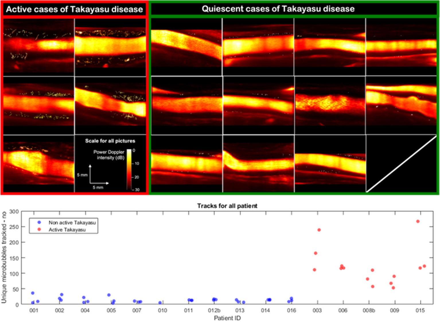
lower panel: microbubble tracking data by individual acquisition for each patient. Patients are presented in groups with active (red) and quiescent (blue) Takayasu arteritis.


Disclosures: G. Goudot, None; A. Jimenez, None; N. Mohamedi, None; j. Sitruk, None; L. Khider, None; H. Mortelette, None; C. Papadacci, None; F. Hyafil, None; M. Tanter, None; E. Messas, None; M. Pernot, None; T. Mirault, None.

To cite this abstract in AMA style:

Goudot G, Jimenez A, Mohamedi N, Sitruk j, Khider L, Mortelette H, Papadacci C, Hyafil F, Tanter M, Messas E, Pernot M, Mirault T. Ultrasound Localization Microscopy as a New Tool for Takayasu’s Arteritis Activity Assessment, a Deep Insight Within Carotid Wall Inflammation [abstract]. Arthritis Rheumatol. 2022; 74 (suppl 9). https://acrabstracts.org/abstract/ultrasound-localization-microscopy-as-a-new-tool-for-takayasus-arteritis-activity-assessment-a-deep-insight-within-carotid-wall-inflammation/. Accessed .
  • Tweet
  • Click to email a link to a friend (Opens in new window) Email
  • Click to print (Opens in new window) Print

« Back to ACR Convergence 2022

ACR Meeting Abstracts - https://acrabstracts.org/abstract/ultrasound-localization-microscopy-as-a-new-tool-for-takayasus-arteritis-activity-assessment-a-deep-insight-within-carotid-wall-inflammation/

Advanced Search

Your Favorites

You can save and print a list of your favorite abstracts during your browser session by clicking the “Favorite” button at the bottom of any abstract. View your favorites »

All abstracts accepted to ACR Convergence are under media embargo once the ACR has notified presenters of their abstract’s acceptance. They may be presented at other meetings or published as manuscripts after this time but should not be discussed in non-scholarly venues or outlets. The following embargo policies are strictly enforced by the ACR.

Accepted abstracts are made available to the public online in advance of the meeting and are published in a special online supplement of our scientific journal, Arthritis & Rheumatology. Information contained in those abstracts may not be released until the abstracts appear online. In an exception to the media embargo, academic institutions, private organizations, and companies with products whose value may be influenced by information contained in an abstract may issue a press release to coincide with the availability of an ACR abstract on the ACR website. However, the ACR continues to require that information that goes beyond that contained in the abstract (e.g., discussion of the abstract done as part of editorial news coverage) is under media embargo until 10:00 AM ET on November 14, 2024. Journalists with access to embargoed information cannot release articles or editorial news coverage before this time. Editorial news coverage is considered original articles/videos developed by employed journalists to report facts, commentary, and subject matter expert quotes in a narrative form using a variety of sources (e.g., research, announcements, press releases, events, etc.).

Violation of this policy may result in the abstract being withdrawn from the meeting and other measures deemed appropriate. Authors are responsible for notifying colleagues, institutions, communications firms, and all other stakeholders related to the development or promotion of the abstract about this policy. If you have questions about the ACR abstract embargo policy, please contact ACR abstracts staff at [email protected].

Wiley

  • Online Journal
  • Privacy Policy
  • Permissions Policies
  • Cookie Preferences

© Copyright 2025 American College of Rheumatology