Session Information
Session Type: Poster Session D
Session Time: 9:00AM-11:00AM
Background/Purpose: Radiography is the most widespread imaging technique used in the evaluation of knee osteoarthritis (KOA), and MRI is known to be specific in characterization of cartilage disease. However, both modalities have their limitations, and there remains need for a reproducible and widely available imaging technique for characterization of KOA disease severity. The utility of ultrasound (US) in inflammatory arthritis is well documented. In KOA, however, the typical sonographic findings are less clear, and can be operator dependent. This study aims to describe sonographic features of KOA, assess reader reliability, and correlate to radiographic findings.
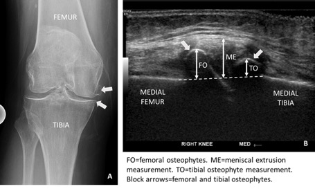
Methods: 161 patients with end stage KOA were prospectively enrolled two weeks prior to knee replacement to undergo knee ultrasound. 31 sonographic features pertinent to KOA were assessed, including osteophyte size, meniscal extrusion (Figure 1), synovial thickness and cartilage erosion. Bangdiwala’s B statistic and intraclass correlation coefficient (ICC) were used to assess rater agreement and interrater reliability, respectively, for 2 raters in 75 knees. Spearman’s rank correlation (ρ) was used to assess for association between US and radiographic findings.
Results: In this cohort of 161 knees, 94 (58%) revealed no to mild suprapatellar synovial thickness. Joint line osteophytes tended to be larger medially, and on the femoral side. The degree of meniscal extrusion was worse at the medial side. There was almost perfect to perfect (0.81-1) rater agreement for 14 of 24 features assessed, including presence of effusion, synovial hyperemia, degree of synovial thickening, patella tendinopathy. Substantial rater agreement (0.70-0.80) was found for medial/lateral joint line synovial thickening, degree of cartilage erosion, presence of Baker’s cyst, and quadriceps tendinosis. Measures of synovial thickness, osteophyte size, degree of meniscal extrusion showed excellent interrater reliability (0.73-0.99), while interrater reliability of Baker’s cyst wall thickness measurement was fair (0.53). Degree of medial and lateral meniscal extrusion correlated with the degree of knee varus and valgus, respectively. Degree of medial and lateral meniscal extrusion, medial tibial and femoral osteophyte size, and lateral femoral osteophyte size correlated with the Kellgren-Lawrence (KL) grade of KOA. However, significant correlations between US and x-ray findings were fair at most (Table 1). Presence or degree of synovial thickening did not correlate with KL grade of KOA, or with the degree of knee varus/valgus.
Conclusion: Patients with KOA had larger medial side osteophytes, and worse degrees of medial meniscal extrusion, compared to the lateral side. The majority of patients presented with no to mild suprapatellar synovial thickening. The majority of sonographic measurements performed in the knee demonstrates high agreement and reliability across raters. Sonographic features of meniscal extrusion and osteophyte size correlated with radiographic measures of knee alignment, and with radiographic K-L grade of KOA. Ongoing studies will correlate these sonographic features of KOA with clinical and histologic findings.
 Figure 1: Radiograph of the right knee (A) with corresponding sonographic image of the medial knee (B) in end stage knee osteoarthritis. Measurement technique for femoral and tibial osteophyte and meniscal extrusion depicted.
 Table 1: Significant correlations between ultrasound and radiographic metrics in patients with end stage knee osteoarthritis.
To cite this abstract in AMA style:
Nwawka O, Lee S, Lin B, Ibrahim S, Brantner C, Sculco P, Parks M, Figgie M, Donlin L, Orange D, Mirza S, Cross M, Rodriguez J, Sculco T, Otero M, Robinson W, Goodman S, Mehta B. Ultrasound in Knee Osteoarthritis: Reader Performance, Sonographic Features, and Correlation with Radiographic Findings [abstract]. Arthritis Rheumatol. 2020; 72 (suppl 10). https://acrabstracts.org/abstract/ultrasound-in-knee-osteoarthritis-reader-performance-sonographic-features-and-correlation-with-radiographic-findings/. Accessed .« Back to ACR Convergence 2020
ACR Meeting Abstracts - https://acrabstracts.org/abstract/ultrasound-in-knee-osteoarthritis-reader-performance-sonographic-features-and-correlation-with-radiographic-findings/
