
Session Information
Date: Sunday, November 13, 2016
Title: Metabolic and Crystal Arthropathies - Poster I: Clinical Practice
Session Type: ACR Poster Session A
Session Time: 9:00AM-11:00AM
Background/Purpose: The utility of ultrasound (US) in aiding the diagnosis of gout has been well established. The latest 2015 gout classification criteria have included the US as one of the criteria. However, in a daily hectic outpatients and inpatients practice, with limited resources, the selection of joints for US examination is crucial. Therefore, we aimed to determine the prevalence of positive US findings in joints of the lower limb among gout patients.
Methods: Data was collected prospectively from 15th January 2016 to 31st May 2016. Patients who fulfilled 2015 ACR/ EULAR gout classification criteria were recruited. US examination of the bilateral first metatarsophalangeal (MTP1), midfoot, ankle and knee joints were performed by an experienced rheumatologist. Typical US lesions in gout such as double contour signs (DCS), tophi, and bone erosions were identified according to the international consensus of OMERACT. The frequencies of the positive US findings were analyzed using a descriptive analysis.
Results: There was a total of 78 patients were recruited, the majority (n=76) were male. The mean age of the patient was 52.3 ¡Ó 16.1 years. A total of 624 joints were examined. The frequency of DCS on ankles, MTP1, knees and midfoot were 98.7%, 96.8%, 95.5%, and 17.9% respectively. The majority of tophi were detected via the US on MTP1 (90.4%), while 9.6% of the midfoot, 11.5% of the ankle, and 16.0% of the knee joints had tophi depositions. Of 156 MTP1 joints examined, 68 (43.6%) had erosions.
Conclusion: MTP1 consistently demonstrated a high frequency of positive US findings (DCS, tophi and bone erosions). The US of the midfoot showed infrequent gout lesions, especially DCS. In daily practice to detect gout, US examinations of the MTP1 are preferred while the US of the midfoot could be omitted. US examination of the ankle and knee joints could be considered in particular looking for DCS. Bone erosions may not be detected by the US if coexistence of tophi depositions on large joint such as knees and ankles.
Table 1. The distribution of double contour signs, tophi, and bone erosions according to joints.
| n (%) | MTP1 | Midfoot |
Ankles |
Knees | ||
|
Anterior |
Medial |
Lateral |
||||
| DCS | 151 (96.8) | 28 (17.9) | 149 (95.5) | 154 (98.7) | 145 (92.9) | 149 (95.5) |
| Tophi | 141 (90.4) | 15 (9.6) | 6 (3.8) | 6 (3.8) | 18 (11.5) | 25 (16.0) |
| Bone erosions | 68 (43.6) | 19 (12.2) |
– |
– |
– |
– |
Abbreviations: DCS, double contour signs; MTP1, first metatarsophalangeal joint.
Figure 1. Typical ultrasound findings on gout patients. 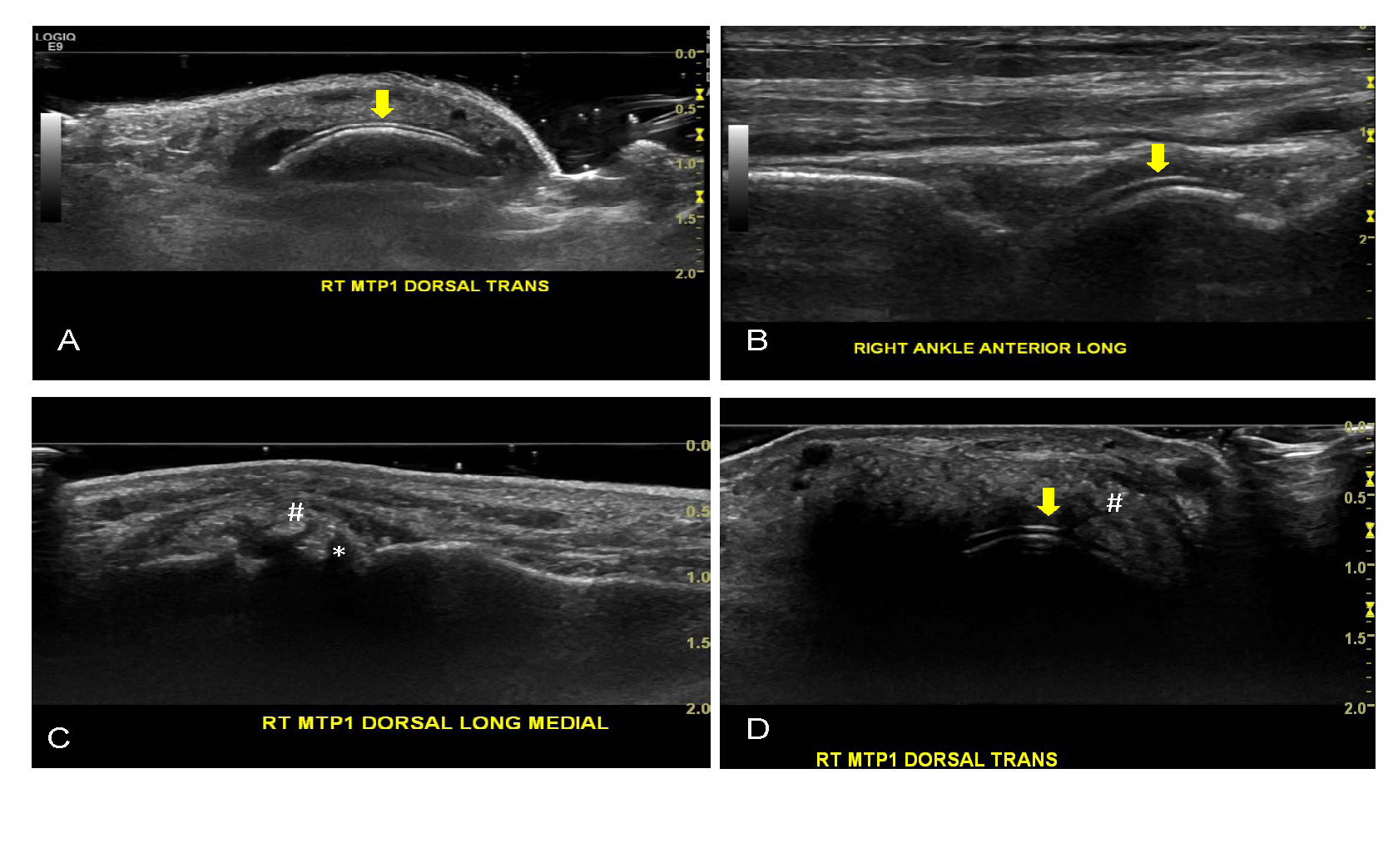
Arrow showed double contour sign. # showed tophi. * showed erosions. Abbreviations: RT, right; MTP1, first metatarsophalangeal joint; TRANS, transverse view.
To cite this abstract in AMA style:
Lin CT, Lim CH, Chen YH, Chen DY. Ultrasound in Gout: The Clinical Application [abstract]. Arthritis Rheumatol. 2016; 68 (suppl 10). https://acrabstracts.org/abstract/ultrasound-in-gout-the-clinical-application/. Accessed .« Back to 2016 ACR/ARHP Annual Meeting
ACR Meeting Abstracts - https://acrabstracts.org/abstract/ultrasound-in-gout-the-clinical-application/