ACR Meeting Abstracts

ACR Meeting Abstracts

  • Meetings
    • ACR Convergence 2024
    • ACR Convergence 2023
    • 2023 ACR/ARP PRSYM
    • ACR Convergence 2022
    • ACR Convergence 2021
    • ACR Convergence 2020
    • 2020 ACR/ARP PRSYM
    • 2019 ACR/ARP Annual Meeting
    • 2018-2009 Meetings
    • Download Abstracts
  • Keyword Index
  • Advanced Search
  • Your Favorites
    • Favorites
    • Login
    • View and print all favorites
    • Clear all your favorites
  • ACR Meetings

Abstract Number: 1664

TRIM21 as a Regulator of UVB-Driven IFN Responses in Lupus

Richard Moore, Gantsetseg Tumurkhuu, Daniel Wallace and Caroline Jefferies, Cedars-Sinai Medical Center, Los Angeles, CA

Meeting: ACR Convergence 2022

Keywords: Animal Model, Cutaneous, interferon, signal transduction, Systemic lupus erythematosus (SLE)

  • Tweet
  • Email
  • Print
Session Information

Date: Monday, November 14, 2022

Title: Abstracts: SLE – Animal Models

Session Type: Abstract Session

Session Time: 10:30AM-11:30AM

Background/Purpose: Patients with systemic lupus erythematosus (SLE) experience photosensitivity where exposure to ultraviolet light B (UVB) drives lupus flares and systemic symptoms. Although the mechanisms linking UVB exposure to these effects are unclear, type I IFNs are known to play a role. TRIM21, an autoantigen in SLE, functions as a negative regulator on the pathways driving IFN expression. Here we explore that the E3 ligase TRIM21 functions to regulate both local and systemic inflammation following UVB exposure in mice and altered expression may explain photosensitivity in SLE.

Methods: C57BL/6 Trim21+/+ (WT) and Trim21-/- (KO) mice were irradiated with UVB (100 mJ/cm2) on shaved dorsal skin with inflammatory responses measured by mRNA transcript levels, histology, and flow cytometry. To determine molecular targets of TRIM21 after UVB exposure, bone marrow-derived macrophages (BMDMs) and murine dermal fibroblasts (MDFs) were isolated from WT and KO mice and treated with the stimulator of IFN genes (STING)-agonist cyclic GMP-AMP (cGAMP) or 100 mJ/cm2 UVB, respectively. Lysates of these cells were used to determine direct effects of intracellular signaling pathways via immunoblotting and immunoprecipitation.

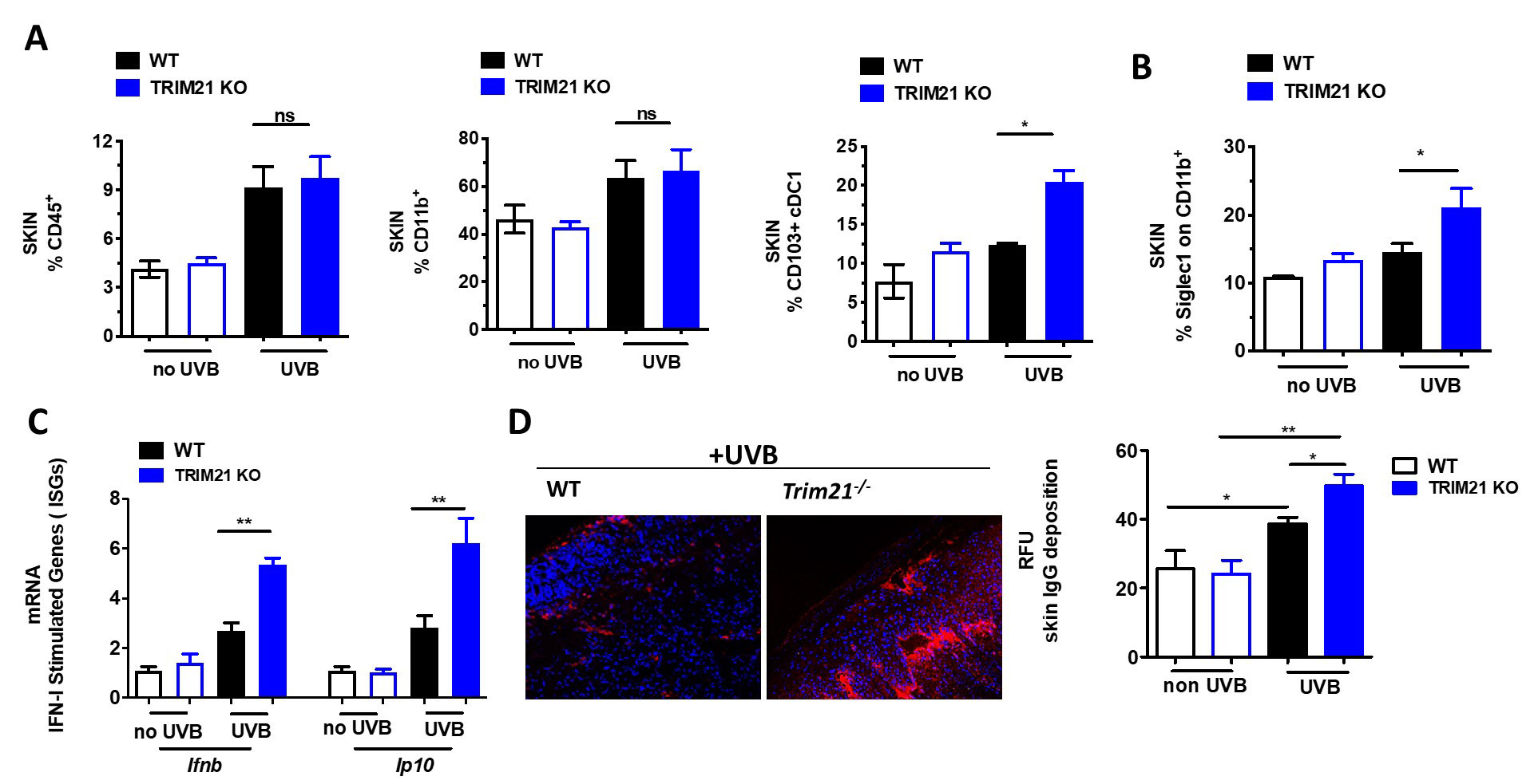
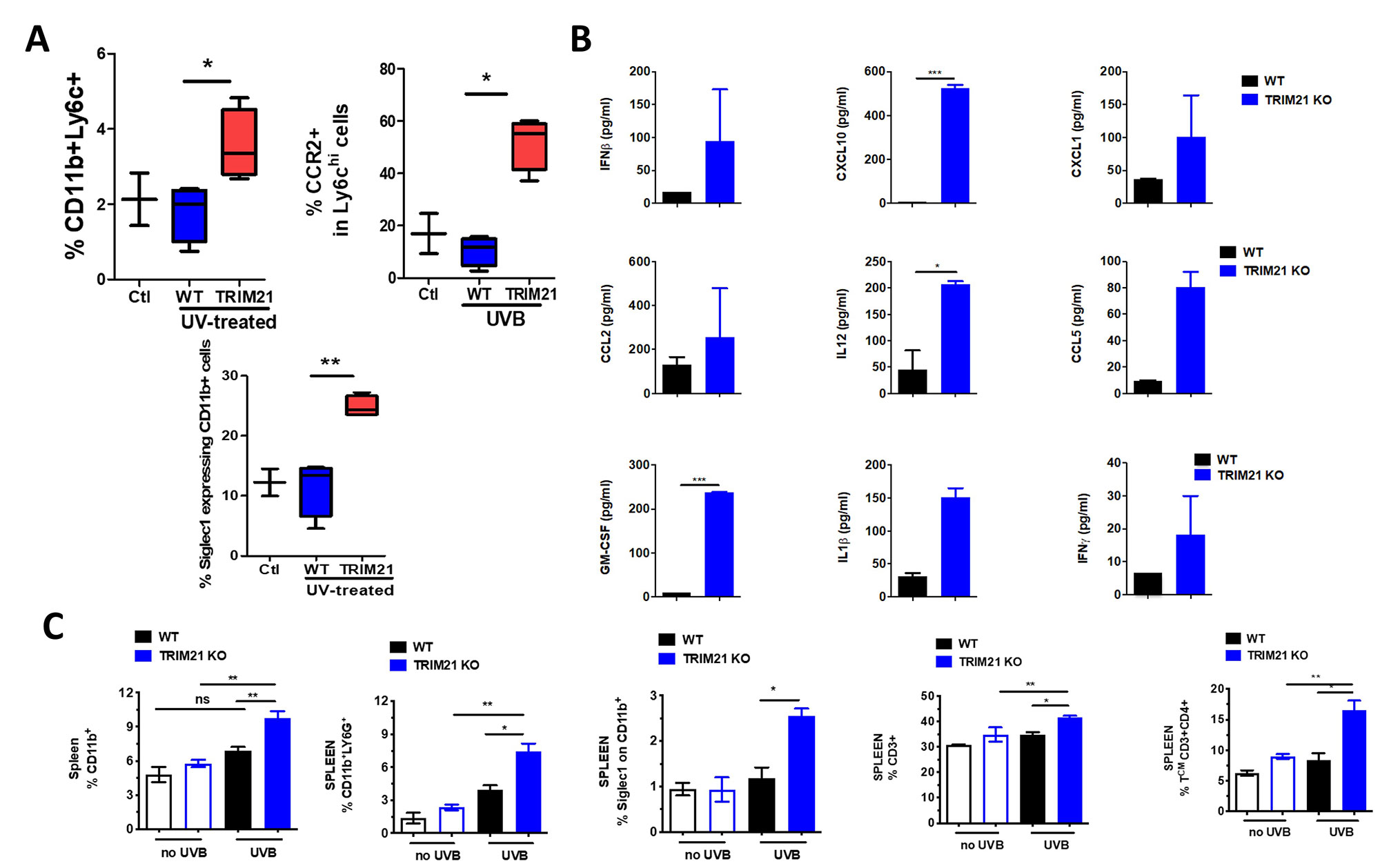
Results: Erythema and edema developed in UVB-exposed skin of both WT and KO mice and associated with increased immune infiltrate within the skin (Fig 1A). However, UVB-exposed skin of KO mice showed increased local levels of conventional dendritic cells, myeloid activation, IFN-stimulated genes (ISGs), and IgG deposition (Fig 1B-D). This response led us to investigate the systemic effects of UVB exposure and indeed, circulating cytokines and activation markers on both circulating monocytes and neutrophils within the spleen were significantly increased (Fig 2A-C). Further, increased levels of both neutrophils and CD4+ T cells were detected in the KO spleen (Fig 2C).

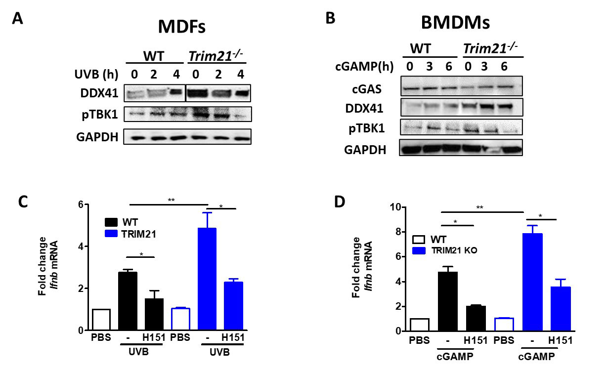
Because we observed local and systemic inflammation, we sought the role TRIM21 has during these processes. To do this, we stimulated MDFs and BMDMs with UVB or cGAS, respectively, and measured protein levels of a known target of TRIM21 E3 ligase activity (DDX41) and phosphorylation of a downstream effector of the STING pathway (TBK1) as well as Ifnb transcript levels. While KO MDFs and BMDMs exhibited increased responses to stimulation, specific inhibition of STING with H151 ameliorated these effects (Fig 3).

Conclusion: In this work, we investigated the effect of Trim21 deficiency in the pathogenesis of the lupus UVB mouse model. Trim21-deficient mice developed heightened local and systemic response to chronic UVB exposure compared to WT mice. Our preliminary analyses confirmed this phenotype is rescued by STING inhibition, keeping with the importance of STING in UVB-driven IFN responses. Taken together, loss of TRIM21 exacerbates UVB-induced skin and systemic inflammation as a result of dysregulated IFN responses in innate immune signaling.

Supporting image 1

Figure 1. C57BL/6 Trim21+/+ (WT) and Trim21-/- (TRIM21 KO) mice were exposed to UVB irradiation. (A) Flow cytometry analysis following the final UVB dose showing the percentage of infiltrating immune cells (CD45+) , myeloid cells (CD11b+), dendritic cells (CD45+CD11C+MHCII+CD103+). (B) The percentage of Siglec1, an IFN-inducible protein, on CD11b+ skin infiltrating cells. (C) Cutaneous expression of mRNA for IFN inducible genes (ISGs) genes following UVB injury, as determined by qPCR. Fold change was calculated against nonirradiated mice, using the 2−ΔΔCt method. Data are compiled from at least 3 independent experiments. (D) IgG deposition in UVB induced skin lesion. A representative pictures and quantification analysis. Values are the mean ± SD. *p<0.05; versus no UV irradiation, One-Way ANOVA with Tukey’s post-hoc test *p<0.05, **p<0.01.

Supporting image 2

Figure 2. C57BL/6 Trim21+/+ (WT) and Trim21-/- (TRIM21 KO) mice were exposed to UVB irradiation. (A) Compared to WT mice, Trim21-/- mice post-UVB exposure had higher circulating monocytes and increased inflammatory markers CCR2 and Siglec1 in Ly6Chi and CD11b+ cells, respectively. (B) Inflammatory cytokines post-UVB determined by Legendflex assay. (C) FACS analysis of spleens following UVB exposure. n = 5-6 per group. All data represent mean ± SD. Statistical significance was determined using One-Way ANOVA with Tukey’s post-hoc test . *p<0.05, **p<0.01, ***p<0.001.

Supporting image 3

Figure 3. Mouse Dermal Fibroblasts (MDFs) and bone marrow derived macrophages (BMDM) derived from WT and Trim21-/- mice were stimulated with UVB or cGAMP for indicated hours. (A-B) The levels of DDX41, cGAS and pTBK1 were determined by immunoblotting. (C-D) The expression of Ifnb mRNA was determined by qPCR. All data represent mean ± SD. Statistical significance was determined using t-test in A; One-Way ANOVA with Tukey’s post-hoc test in B . *p<0.05, **p<0.01, ***p<0.001.


Disclosures: R. Moore, None; G. Tumurkhuu, None; D. Wallace, None; C. Jefferies, None.

To cite this abstract in AMA style:

Moore R, Tumurkhuu G, Wallace D, Jefferies C. TRIM21 as a Regulator of UVB-Driven IFN Responses in Lupus [abstract]. Arthritis Rheumatol. 2022; 74 (suppl 9). https://acrabstracts.org/abstract/trim21-as-a-regulator-of-uvb-driven-ifn-responses-in-lupus/. Accessed .
  • Tweet
  • Email
  • Print

« Back to ACR Convergence 2022

ACR Meeting Abstracts - https://acrabstracts.org/abstract/trim21-as-a-regulator-of-uvb-driven-ifn-responses-in-lupus/

Advanced Search

Your Favorites

You can save and print a list of your favorite abstracts during your browser session by clicking the “Favorite” button at the bottom of any abstract. View your favorites »

All abstracts accepted to ACR Convergence are under media embargo once the ACR has notified presenters of their abstract’s acceptance. They may be presented at other meetings or published as manuscripts after this time but should not be discussed in non-scholarly venues or outlets. The following embargo policies are strictly enforced by the ACR.

Accepted abstracts are made available to the public online in advance of the meeting and are published in a special online supplement of our scientific journal, Arthritis & Rheumatology. Information contained in those abstracts may not be released until the abstracts appear online. In an exception to the media embargo, academic institutions, private organizations, and companies with products whose value may be influenced by information contained in an abstract may issue a press release to coincide with the availability of an ACR abstract on the ACR website. However, the ACR continues to require that information that goes beyond that contained in the abstract (e.g., discussion of the abstract done as part of editorial news coverage) is under media embargo until 10:00 AM ET on November 14, 2024. Journalists with access to embargoed information cannot release articles or editorial news coverage before this time. Editorial news coverage is considered original articles/videos developed by employed journalists to report facts, commentary, and subject matter expert quotes in a narrative form using a variety of sources (e.g., research, announcements, press releases, events, etc.).

Violation of this policy may result in the abstract being withdrawn from the meeting and other measures deemed appropriate. Authors are responsible for notifying colleagues, institutions, communications firms, and all other stakeholders related to the development or promotion of the abstract about this policy. If you have questions about the ACR abstract embargo policy, please contact ACR abstracts staff at [email protected].

Wiley

  • Online Journal
  • Privacy Policy
  • Permissions Policies
  • Cookie Preferences

© Copyright 2025 American College of Rheumatology