ACR Meeting Abstracts

ACR Meeting Abstracts

  • Meetings
    • ACR Convergence 2024
    • ACR Convergence 2023
    • 2023 ACR/ARP PRSYM
    • ACR Convergence 2022
    • ACR Convergence 2021
    • ACR Convergence 2020
    • 2020 ACR/ARP PRSYM
    • 2019 ACR/ARP Annual Meeting
    • 2018-2009 Meetings
    • Download Abstracts
  • Keyword Index
  • Advanced Search
  • Your Favorites
    • Favorites
    • Login
    • View and print all favorites
    • Clear all your favorites
  • ACR Meetings

Abstract Number: 256

Tracking Digital Ulcers in Systemic Sclerosis: Feasibility Study Assessing Lesion Area from Patient-Recorded Smartphone Photographs

Graham Dinsdale1, Tonia Moore2, Joanne Manning2, Andrea Murray1, Ross Atkinson3, Karen Ousey4, Mark Dickinson5, Christopher Taylor6 and Ariane L. Herrick1,7, 1Division of Musculoskeletal & Dermatological Sciences, University of Manchester, Salford Royal Hospital NHS Foundation Trust, Manchester Academic Health Science Centre, Manchester, UK, Manchester, United Kingdom, 2Salford Royal Hospital NHS Foundation Trust, Salford, UK, Salford, United Kingdom, 3School of Health Sciences, Division of Nursing, Midwifery & Social Work, University of Manchester, Manchester, UK, Manchester, United Kingdom, 4Division of Podiatry and Clinical Sciences, University of Huddersfield, Huddersfield, UK, Huddersfield, United Kingdom, 5Photon Science Institute, School of Physics and Astronomy, University of Manchester, Manchester, UK, Manchester, United Kingdom, 6Centre for Imaging Sciences, Division of Informatics, Imaging & Data Sciences, University of Manchester, Manchester, UK, Manchester, United Kingdom, 7NIHR Manchester Musculoskeletal Biomedical Research Unit, Central Manchester NHS Foundation Trust, Manchester Academic Health Science Centre, Manchester, UK, Manchester, United Kingdom

Meeting: 2017 ACR/ARHP Annual Meeting

Date of first publication: September 18, 2017

Keywords: Imaging, imaging techniques, systemic sclerosis and ulcers

  • Tweet
  • Email
  • Print
Session Information

Date: Sunday, November 5, 2017

Title: Imaging of Rheumatic Diseases Poster I

Session Type: ACR Poster Session A

Session Time: 9:00AM-11:00AM

Background/Purpose: Approximately 50% of patients with systemic sclerosis (SSc) will develop painful digital (finger) ulcers (DUs) at some point during their disease course. These lesions can be extremely disabling and are often refractory to treatment, requiring close monitoring of healing progression. Also, DUs are often the primary outcome measure in clinical trials of SSc-related digital vasculopathy. Our aims were to: (1) demonstrate the feasibility of patients with SSc taking smartphone photographs of their DUs, and (2) use software image analysis on collected images to track lesion area as a marker of wound status.

Methods: Patients with SSc and incident DUs were asked to photograph their lesion(s), using their own smartphone, once per day for a maximum period of 35 days. Patients received normal clinical wound care for the duration of the study, which in most cases was patient self-management. Image length scales were initially calibrated using a fixed (non-varying) object in each image sequence, allowing relative length/area tracking throughout a sequence. Using digital planimetry software (developed and successfully tested in previous work on digital ulcer photographs [1]), lesion area was measured by fitting an elliptical shape to the wound image by a single observer. Areas from each image were then normalised to the area measured first in the time sequence.

Results: Image sequences describing 7 lesions were collected in-person at the end of the study period from 4 patients. The 7 image sequences cover median [range] duration of 29 [13-35] days. The relative area time course for each lesion is shown in Figure 1. At sequence end, relative lesion areas had, on average, reduced to 56% of the area measured on day 1, with 6 out of 7 lesions reducing in size over the time course.

Conclusion: We have demonstrated the feasibility of patients with SSc collecting images of digital ulcers using smartphone cameras. Images collected are of sufficient quality to allow software monitoring of wound progression (healing or worsening). Further work to build a smartphone app for lesion monitoring (for use in both clinical practice and as an outcome measure in clinical trials) is now required.

References

1.       Simpson et al. Arthritis Care & Research, 2017, doi:10.1002/acr.23300

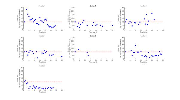
Figure 1. Normalised lesion area time course plots (blue squares) for each of seven digital ulcers. Red dashed line equals 100% relative area, i.e. equal to the value at the start of the sequence. Only lesion 3 showed an increase in area over the course of the study.


Disclosure: G. Dinsdale, None; T. Moore, None; J. Manning, None; A. Murray, None; R. Atkinson, None; K. Ousey, None; M. Dickinson, None; C. Taylor, None; A. L. Herrick, None.

To cite this abstract in AMA style:

Dinsdale G, Moore T, Manning J, Murray A, Atkinson R, Ousey K, Dickinson M, Taylor C, Herrick AL. Tracking Digital Ulcers in Systemic Sclerosis: Feasibility Study Assessing Lesion Area from Patient-Recorded Smartphone Photographs [abstract]. Arthritis Rheumatol. 2017; 69 (suppl 10). https://acrabstracts.org/abstract/tracking-digital-ulcers-in-systemic-sclerosis-feasibility-study-assessing-lesion-area-from-patient-recorded-smartphone-photographs/. Accessed .
  • Tweet
  • Email
  • Print

« Back to 2017 ACR/ARHP Annual Meeting

ACR Meeting Abstracts - https://acrabstracts.org/abstract/tracking-digital-ulcers-in-systemic-sclerosis-feasibility-study-assessing-lesion-area-from-patient-recorded-smartphone-photographs/

Advanced Search

Your Favorites

You can save and print a list of your favorite abstracts during your browser session by clicking the “Favorite” button at the bottom of any abstract. View your favorites »

All abstracts accepted to ACR Convergence are under media embargo once the ACR has notified presenters of their abstract’s acceptance. They may be presented at other meetings or published as manuscripts after this time but should not be discussed in non-scholarly venues or outlets. The following embargo policies are strictly enforced by the ACR.

Accepted abstracts are made available to the public online in advance of the meeting and are published in a special online supplement of our scientific journal, Arthritis & Rheumatology. Information contained in those abstracts may not be released until the abstracts appear online. In an exception to the media embargo, academic institutions, private organizations, and companies with products whose value may be influenced by information contained in an abstract may issue a press release to coincide with the availability of an ACR abstract on the ACR website. However, the ACR continues to require that information that goes beyond that contained in the abstract (e.g., discussion of the abstract done as part of editorial news coverage) is under media embargo until 10:00 AM ET on November 14, 2024. Journalists with access to embargoed information cannot release articles or editorial news coverage before this time. Editorial news coverage is considered original articles/videos developed by employed journalists to report facts, commentary, and subject matter expert quotes in a narrative form using a variety of sources (e.g., research, announcements, press releases, events, etc.).

Violation of this policy may result in the abstract being withdrawn from the meeting and other measures deemed appropriate. Authors are responsible for notifying colleagues, institutions, communications firms, and all other stakeholders related to the development or promotion of the abstract about this policy. If you have questions about the ACR abstract embargo policy, please contact ACR abstracts staff at [email protected].

Wiley

  • Online Journal
  • Privacy Policy
  • Permissions Policies
  • Cookie Preferences

© Copyright 2025 American College of Rheumatology