Session Information
Session Type: Poster Session B
Session Time: 9:00AM-11:00AM
Background/Purpose: Serum urate (SU) lowering has been reported to improve gout erosions. We observed that it may also lead to compaction of toe MTP or IP joints involved by severe gouty arthropathy.
Methods: We performed a retrospective analysis of 10 patients with severe tophaceous gout and destructive arthropathy, who did not receive urate-lowering drugs (ULDs) before inclusion and developed toe shortening (a sign we called the shrinking toe sign) during urate lowering treatment (ULT) at the Vien Gut Medical Center (Ho Chi Minh City, Vietnam). Data were retrieved from anonymized electronic files, including sequential foot radiographs and photographs. Antero-posterior non-weight bearing digital radiographs of the feet taken before ULT start, and during follow-up, were analyzed by using the COREL DRAW software (Corel Corporation, CANADA), which allowed standardization of the size and orientation of patients’ radiographs, and measurement of toe shortenings.
Results: The 10 patients were men with a mean age of 39 +/- 6.6 years at gout onset. At inclusion, their mean age was 51.4 (SD 9.6) years, mean gout duration 12.4 (3.8) years, mean uricemia 528.2 (51.9) micromoles/l, mean BMI 22.5 (3.8) kg/m2. Patients were treated by progressively increased doses of allopurinol (n=6; mean maintenance dose: 650 mg/d; range 450-900), or febuxostat (n=4; mean maintenance dose 130 mg/d; range 80-160). All patients achieved the < 300 micromoles/l serum urate (SU) target after a mean of 7.4 months (range 1-15).
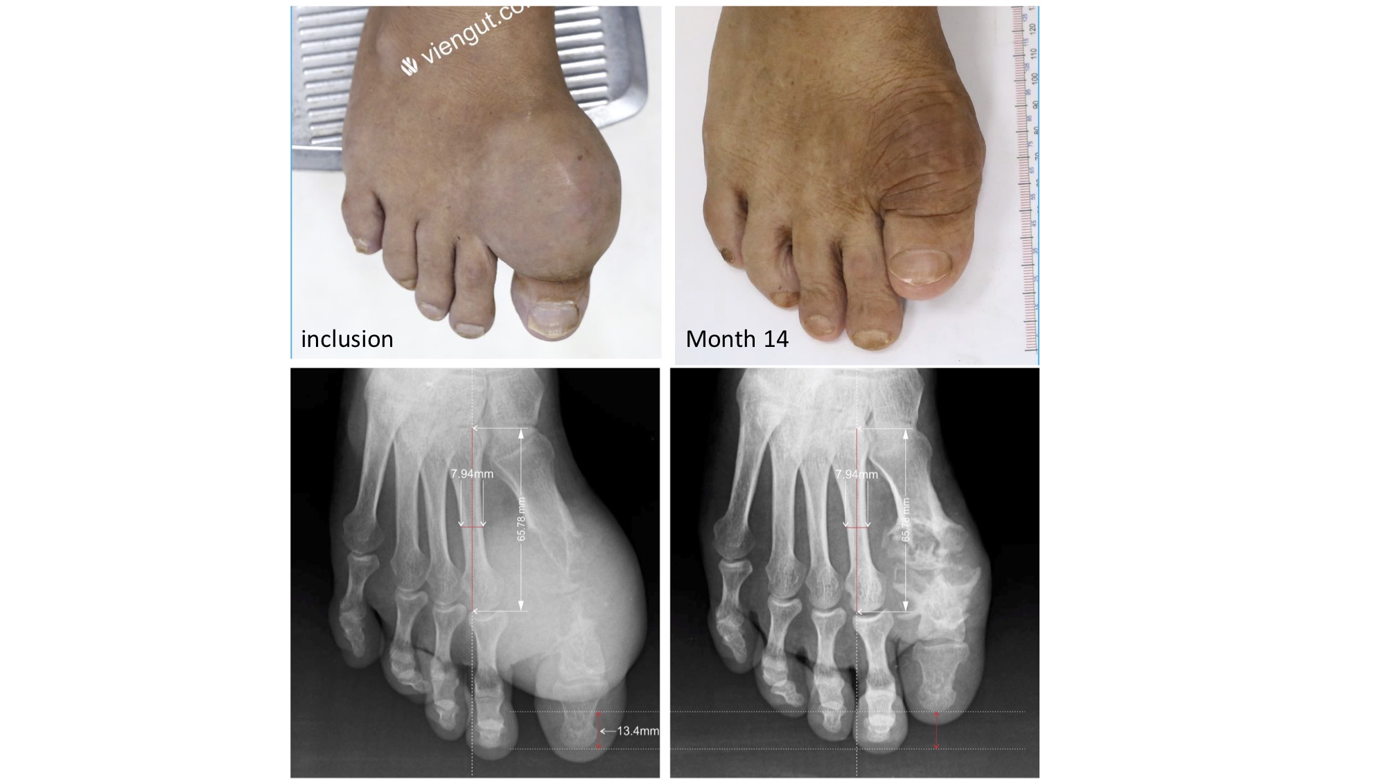
During a mean total radiological follow-up of 27 (15) months (range 6-48), the 10 patients had a mean of 1.8 radiographs each (range 1-4). The shrinking toe sign involved 12 toes: 7 first MTPs (Fig.) (mean shortening 8.7 mm; range: 3-13.8), 3 second MTPs ( mean 12.4 mm; range: 9.5-17.9), and 2 second toe DIPs (9.5 and 4.4 mm). Toe shortening was also observed clinically, as confirmed by sequential photographs (Fig.).
In one patient, subchondral lytic bone collapse of P1 basis was observed 9 months before the SU target was reached, leading to shortening of a big toe, with no sign of tophus decrease, suggesting that the shrinking toe sign can occur in poorly treated gout. In the other 9 patients, shrinking toes were observed after the target had been reached at a mean follow-up of 22.9 months (range 2-47). Dissolution of MSU deposits within the joint space and/or subchondral bone lytic areas, leading to their collapse, despite the frequent observation of bone reconstruction, appeared to be the main explanation for toe shortening, associated with subluxation of the involved joint in 3 patients. 5 patients underwent multiple follow-up radiographs; in 4, toes shortened progressively with time in parallel with decrease of tophus opacities, whereas the 5th, who had 3 follow-up radiographs up to 27 months, exhibited maximum shortening at the first follow-up radiograph (month 11), when tophus opacities had already vanished.
Conclusion: The shrinking toe sign that we described in this study involved the 1st and 2d toes, where constraints are maximized. It may be a feature of severe gouty arthropathy, but was mainly observed during MSU dissolution under ULT, which appeared to fragilize the involved joints.
 Example of the shrinking toe sign
To cite this abstract in AMA style:
Bardin T, Quang N, Nghia H, Khoi T, EA H, Bousson V, Richette P. The Shrinking Toe: A Sign of Crystal Dissolution During Urate Lowering Treatment of Severe Gouty Arthropathy [abstract]. Arthritis Rheumatol. 2020; 72 (suppl 10). https://acrabstracts.org/abstract/the-shrinking-toe-a-sign-of-crystal-dissolution-during-urate-lowering-treatment-of-severe-gouty-arthropathy/. Accessed .« Back to ACR Convergence 2020
ACR Meeting Abstracts - https://acrabstracts.org/abstract/the-shrinking-toe-a-sign-of-crystal-dissolution-during-urate-lowering-treatment-of-severe-gouty-arthropathy/
