Session Information
Date: Sunday, November 13, 2016
Title: Rheumatoid Arthritis – Clinical Aspects - Poster I: Clinical Characteristics/Presentation/Prognosis
Session Type: ACR Poster Session A
Session Time: 9:00AM-11:00AM
Background/Purpose: For patients (pts) with rheumatoid arthritis (RA), quantification of radiographic progression, through measures such as modified total Sharp score (mTSS), is not routinely captured in daily clinical practice. Surrogate markers of radiographic progression may facilitate identification of pts who require closer monitoring. The aim was to assess the relationship between elevations in C-reactive protein (CRP) values, over 10 years (yrs), with physical function and radiographic progression.
Methods: This post hoc analysis used data from 2 trials: PREMIER,1a 2-yr randomized, controlled trial (RCT) in methotrexate (MTX)-naïve, early RA pts, receiving MTX, adalimumab (ADA) or ADA+MTX, and DE019,2a 1-yr RCT in RA pts with an inadequate response to MTX, receiving Placebo or ADA on background MTX. In both trials, pts completing the RCT could enter an open label extension (OLE) for a total treatment period of 10 yrs. CRP and the disability index of the health assessment questionnaire (HAQ-DI) were collected at regular visits throughout the RCTs and OLEs. Radiographic data were collected at baseline, 6 months, 1, 2, 3, 5, 6, 8 and 10 yrs. Spearman coefficients assessing correlations between CRP elevations (defined as CRP >upper limit of normal [ULN]) and HAQ-DI were calculated at all visits. The association of radiographic progression (ΔmTSS ≥0.5) with CRP elevations and time-averaged (TA)-CRP was assessed between x-ray reads by logistic regression and descriptive statistics.
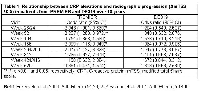
Results: In both PREMIER and DE019, a significant, positive correlation was observed between CRP elevations and HAQ-DI at nearly all time points during the RCTs, and approximately half of the time points throughout the OLEs. Further, CRP elevations were associated with radiographic progression during the first half of PREMIER but these associations were not observed at any time point in DE019 (Table 1). For pts who experienced ≥1 CRP elevation >ULN between x-ray reads, TA-CRP was significantly associated with radiographic progression only at yrs 5 and 6 in PREMIER. Radiographic progression was observed frequently among pts with elevations in CRP between x-ray reads; however, radiographic progression was still seen in 20-52% of pts without CRP elevations. Pts receiving MTX monotherapy in both RCTs experienced an increase in CRP elevations compared to those pts receiving ADA+MTX (91% vs 55% for MTX and ADA+MTX, respectively, at 2 yrs in PREMIER; 68% vs 33% for MTX and ADA+MTX, respectively, at 1 yr in DE019). Consistently, treatment was significantly associated with radiographic progression, as assessed by logistic regression.
Conclusion: CRP is an important marker of inflammation that can be used to identify pts at risk of radiographic progression, and this relationship appears dependent on pt disease characteristics. Structural evaluation remains important until further predictors are identified.
To cite this abstract in AMA style:
Kavanaugh A, Haraoui B, Sunkureddi P, Wolfe B, Wang L, Suboticki J, Keystone E. The Relationship Between Elevations in CRP with Physical Function and Radiographic Progression over the Long-Term in Patients with Rheumatoid Arthritis [abstract]. Arthritis Rheumatol. 2016; 68 (suppl 10). https://acrabstracts.org/abstract/the-relationship-between-elevations-in-crp-with-physical-function-and-radiographic-progression-over-the-long-term-in-patients-with-rheumatoid-arthritis/. Accessed .« Back to 2016 ACR/ARHP Annual Meeting
ACR Meeting Abstracts - https://acrabstracts.org/abstract/the-relationship-between-elevations-in-crp-with-physical-function-and-radiographic-progression-over-the-long-term-in-patients-with-rheumatoid-arthritis/

