Session Information
Date: Sunday, November 17, 2024
Title: Innate Immunity Poster
Session Type: Poster Session B
Session Time: 10:30AM-12:30PM
Background/Purpose: Previous in vitro investigations done by our group into the utility of CB2R activation to treat dermatomyositis (DM) used stimulants that activated pathways not specifically relevant to DM. Here, we tested CB2R activation to determine its anti-inflammatory effects on pathways biologically relevant to DM.
Methods: We collected CB2R positivity data on 3 Amyopathic DM patients and 3 Classic DM patients and collected cytokine data on 4 Amyopathic DM patients and 3 Classic DM patients. CB2R positivity data was obtained by analyzing patient PBMCs via flow cytometry. IFNβ was used as a marker for inflammation to test CB2R activation and it was measured in cells stimulated by the following: dsRNA for RIG1, dsDNA for cGAS, LPS for TLR4, and LPS/ATP for NLRP3. The CB2R agonist JWH133 was used to pretreat PBMCs before stimulation. The resulting PBMCs were stained, and flow cytometry data was acquired using FACS Symphony A3 Lite and analyzed with FlowJo V10.7.1. Statistical analysis was performed using GraphPad Prism V9. Comparisons between Amyopathic and Classic DM CB2R positivity and IFNB positivity were made with Student’s t-test.
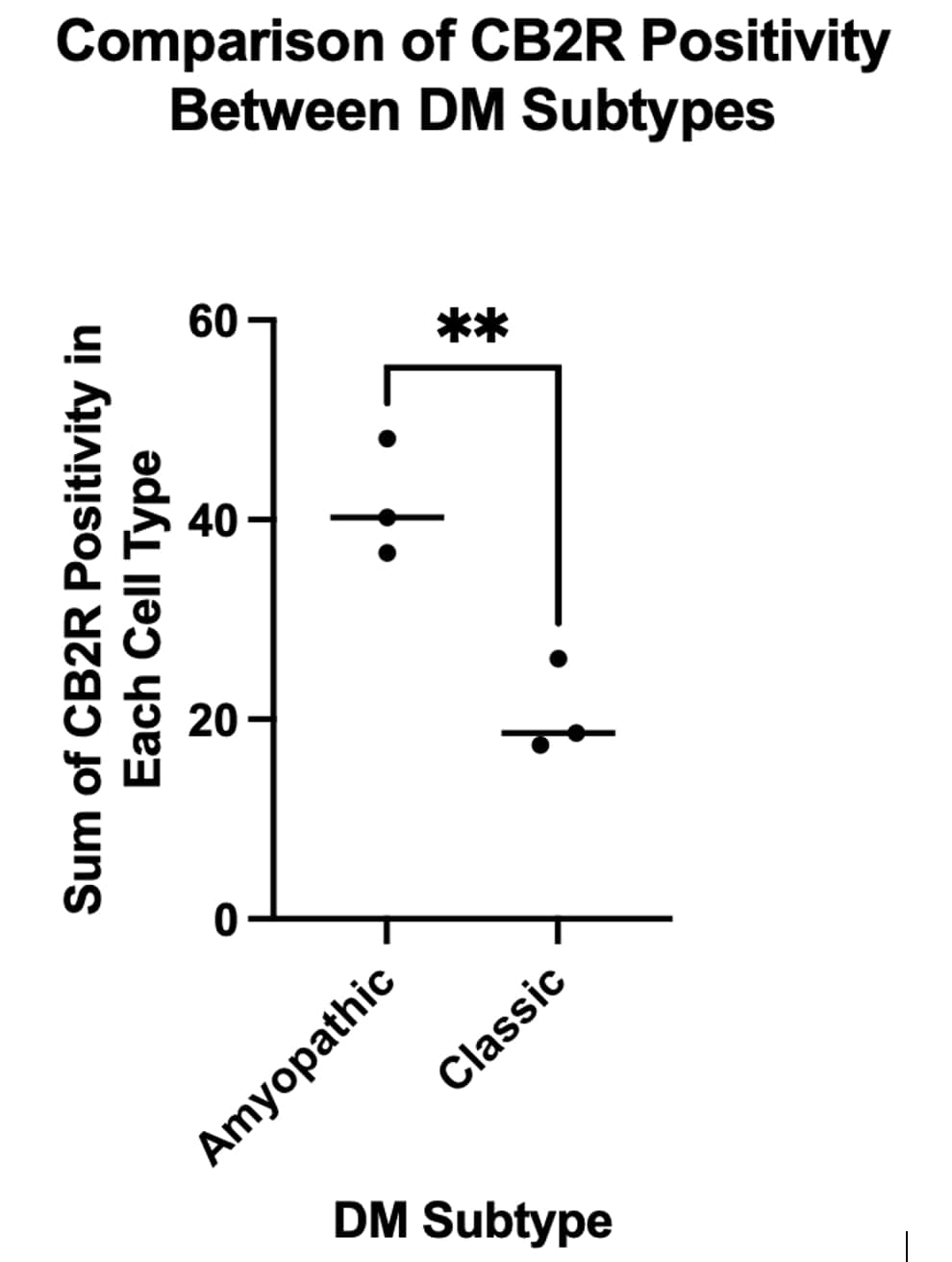
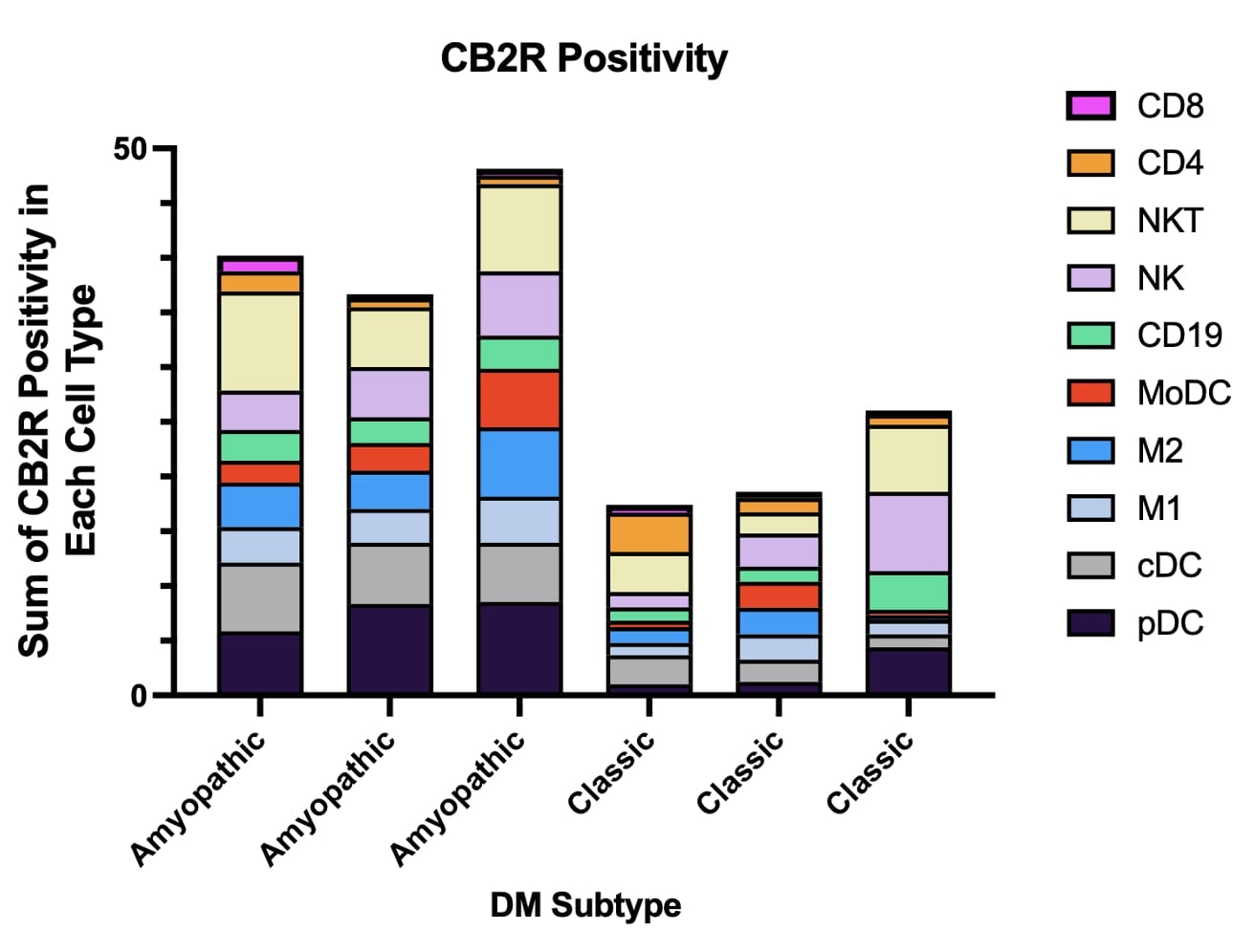
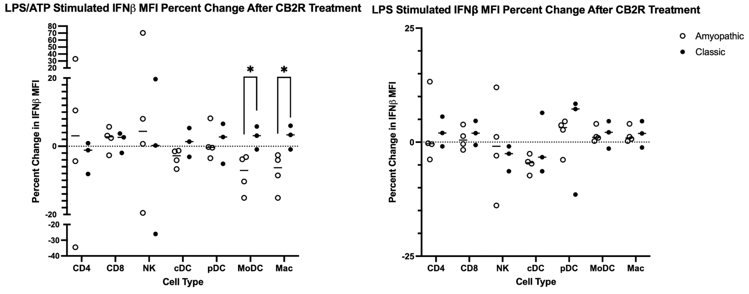
Results: CB2R positivity data was collected for PBMC CB2R Frequency of Parent (FoP) for the following cell types: CD4, CD8, NKT, NK, CD19, MoDC, M2, M1, cDC, and pDC. CB2R positivity was compared by summing the CB2R FoP for the 10 investigated cell types. Amyopathic DM PBMCS were found to be 101.3% more positive for CB2R compared to Classic DM PBMCS (p=0.0085) (Figure 1). For Amyopathic DM PBMCs, the top five cell types accounting for CB2R positivity in decreasing order are the following: pDC, NKT, cDC, NK, M2, and M1 (Figure 2). For Classic DM PBMCs, the top five cell types accounting for CB2R positivity in decreasing order are the following: NK, NKT, pDC, CD19, and cDC (Figure 2). In amyopathic DM PBMCs stimulated by LPS/ATP to target the NLRP3 inflammasome, CB2R activation resulted in a significant reduction in IFNβ MFI for MoDCs (p=0.0397) and Macs (p=0.0457) with a similar trend was observed in cDCs relative to classic DM PBMCS (Figure 3). On the other hand, no difference in IFNβ response to CB2R activation was observed across all cell types investigated between classic and amyopathic DM PBMCs stimulated with LPS only to target TLR4 (Figure 3).
Conclusion: Amyopathic DM PBMCs are significantly more positive for CB2R. CB2R activation appears to be more effective in reducing NLRP3-induced IFNβ production in amyopathic DM MoDCs and Macs compared to classic DM MoDCs and Macs. In contrast, TLR4-stimulated amyopathic DM PBMCs treated with CB2RA show no reduction in IFNβ, suggesting that CB2R activation may be more effective in reducing NLRP3-induced inflammation compared to TLR4-induced inflammation.
To cite this abstract in AMA style:
Dhiman R, Eldaboush A, Vijayarangan N, Kang D, Stone C, Kodali N, Diaz D, Werth V. The Effects of CB2R Activation on Inflammatory Pathways in Dermatomyositis [abstract]. Arthritis Rheumatol. 2024; 76 (suppl 9). https://acrabstracts.org/abstract/the-effects-of-cb2r-activation-on-inflammatory-pathways-in-dermatomyositis/. Accessed .« Back to ACR Convergence 2024
ACR Meeting Abstracts - https://acrabstracts.org/abstract/the-effects-of-cb2r-activation-on-inflammatory-pathways-in-dermatomyositis/


