
Session Information
Session Type: Abstract Submissions (ACR)
Background/Purpose: Syndesmophytes, the hallmark of structural damage in ankylosing spondylitis (AS), are typically visualized 2-dimensionally by radiographs. However, these projections only allow a limited view of syndesmophytes and their locations along the vertebral rim. With computed tomography (CT), a clear and complete view of syndesmophytes and their precise location is possible. The question immediately arises whether the distribution of syndesmophytes along the vertebral rim is random, as might be suspected if due to inflammation alone, or if they occur more commonly at particular locations. The radial distribution of syndesmophytes along the vertebral rim, if not random, could provide clues about the drivers of syndesmophyte development.
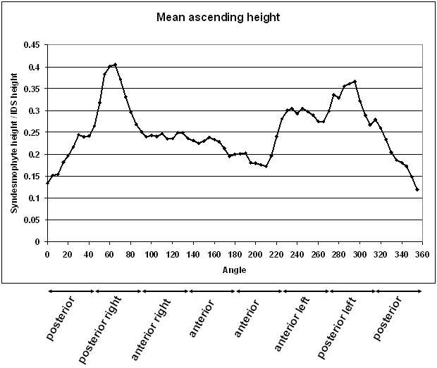
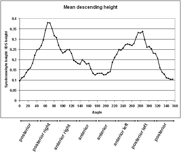
Methods: We scanned 38 patients (31 men and 7 women, mean age of 46.1 years) at the thoracolumbar junction (T10 to L4) using CT, providing 4 intervertebral disk spaces (IDS) per patient for analysis. Syndesmophytes were identified by a computer algorithm that detects them as bone projecting from the periphery of vertebral endplates. We divided the vertebral rim into angular sectors and measured syndesmophyte height every five degrees. We standardized syndesmophyte height by dividing by the height of the disk so that a value of 1 indicated bridging. We plotted radial heights for both ascending and descending syndesmophytes. Bridged syndesmophytes were counted as both ascending and descending. We used the permutation testing for correlated angular data devised by Follmann and Proschan to assess whether the distribution around the rim was uniform or not.
Results: Of 152 IDSs processed, 95 had syndesmophytes. For both ascending and descending syndesmophytes, the mean height distribution along the vertebral rim was non-random (p<.0001 for both). The mean radial height plots show that the most common location for syndesmophytes was posterolateral (Figure). The second most common location was anterior left. Ascending syndesmophytes were more frequent and taller than descending syndesmophytes. In a separate analysis, we sorted the 95 IDSs in rank order of increasing fusion. Isolated syndesmophytes and small (early) syndesmophytes were most common in the posterolateral left and posterolateral right locations, suggesting that syndesmophyte growth most often begins at these locations.
Conclusion: The distribution of syndesmophytes along the vertebral rim is strongly non-random, with posterolateral the most common location. The non-random distribution suggests that mechanical factors may be a driver of syndesmophyte growth.
.
Disclosure:
S. Tan,
None;
A. Dasgupta,
None;
J. Yao,
None;
J. A. Flynn,
None;
L. Yao,
None;
M. M. Ward,
None.
« Back to 2013 ACR/ARHP Annual Meeting
ACR Meeting Abstracts - https://acrabstracts.org/abstract/syndesmophyte-distribution-along-the-vertebral-rim-in-ankylosing-spondylitis-is-non-random/