ACR Meeting Abstracts

ACR Meeting Abstracts

  • Meetings
    • ACR Convergence 2024
    • ACR Convergence 2023
    • 2023 ACR/ARP PRSYM
    • ACR Convergence 2022
    • ACR Convergence 2021
    • ACR Convergence 2020
    • 2020 ACR/ARP PRSYM
    • 2019 ACR/ARP Annual Meeting
    • 2018-2009 Meetings
    • Download Abstracts
  • Keyword Index
  • Advanced Search
  • Your Favorites
    • Favorites
    • Login
    • View and print all favorites
    • Clear all your favorites
  • ACR Meetings

Abstract Number: 1067

Spontaneous Aggregation of the Anti-Viral Mavs Protein in Certain Systemic Lupus Erythematosus Patients May Explain Excessive Type I Interferon Production

Philip L. Cohen1 and Wen-Hai Shao2, 1Rheumatology, Temple University, Philadelphia, PA, 2Medicine, Temple University School of Medicine, Philadelphia, PA

Meeting: 2012 ACR/ARHP Annual Meeting

Keywords: interferons, lupus nephritis and viruses

  • Tweet
  • Click to email a link to a friend (Opens in new window) Email
  • Click to print (Opens in new window) Print
Session Information

Title: Innate Immunity and Rheumatic Disease

Session Type: Abstract Submissions (ACR)

Background/Purpose: Patients with systemic lupus (SLE) often have increased type I interferon levels (IFN-I) and activation of IFN-inducible genes (IFN signature).  The mitochondrial adaptor protein MAVS (also known as IPS1, VISA or CARDIF) is a key intermediary in the RIG-I pathway, where viral RNA triggers a conformational change in RIG-I, leading to MAVS activation and then downstream activation of IKK and TBK1, with subsequent IFN production driven by IRF-3/7 (IRF3 for IFN-beta; IRF7 for IFN-alpha) and NFkB activation and translocation.  Using in vitro methods, it has been observed that MAVS may form large prion-like aggregates that might stimulate IFN-I activation in a potent and prolonged fashion (Hou et. al., Cell 146:448, 2011).  We wondered if such aggregates might be detectable ex vivo in SLE patients, and whether they might play a role in the sustained increased production of IFN-I.

Methods: Peripheral blood mononuclear cells (PBMCs) were isolated from 17 patients fulfilling ACR criteria for SLE, and from 9 controls. Thirty million PBMCs were lysed and supernatants loaded onto semi-denaturing 1.5% vertical agarose gels.  After electrophoresis, the proteins were transferred to membranes for immunoblotting with anti-MAVS antibody or anti-beta-actin.

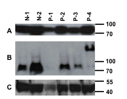
Results: Four of 17 SLE patients showed clear MAVS aggregation, with essentially all of their MAVS protein in a high molecular weight aggregated form.  None of 9 controls had abnormal MAVS. Three of the four aggregation-positive SLE patients had nephritis and the fourth had lung involvement.  SLEDAI scores of MAVS-aggregate positive SLE patients did not differ from patients with normal molecular weight MAVS.  Patient 4 (P-4) shows the aggregated MAVS phenotype in the western blot below (Panel B, P-4).  Denatured MAVS immunoblotting is shown in panel A and actin immunoblotting in panel C.  N-1 and N-2 are normal controls.  P-1 has less protein loaded and no MAVS band is discernible.

Conclusion: This is the first report of aggregated MAVS in human cells.  The significance of this abnormality needs further investigation, it is possible that prolonged and increased IFN-I production could result from such MAVS aggregation, and that the poorly degradable prion-like protein could signal IFN-I production for prolonged periods.


Disclosure:

P. L. Cohen,
None;

W. H. Shao,
None.

  • Tweet
  • Click to email a link to a friend (Opens in new window) Email
  • Click to print (Opens in new window) Print

« Back to 2012 ACR/ARHP Annual Meeting

ACR Meeting Abstracts - https://acrabstracts.org/abstract/spontaneous-aggregation-of-the-anti-viral-mavs-protein-in-certain-systemic-lupus-erythematosus-patients-may-explain-excessive-type-i-interferon-production/

Advanced Search

Your Favorites

You can save and print a list of your favorite abstracts during your browser session by clicking the “Favorite” button at the bottom of any abstract. View your favorites »

All abstracts accepted to ACR Convergence are under media embargo once the ACR has notified presenters of their abstract’s acceptance. They may be presented at other meetings or published as manuscripts after this time but should not be discussed in non-scholarly venues or outlets. The following embargo policies are strictly enforced by the ACR.

Accepted abstracts are made available to the public online in advance of the meeting and are published in a special online supplement of our scientific journal, Arthritis & Rheumatology. Information contained in those abstracts may not be released until the abstracts appear online. In an exception to the media embargo, academic institutions, private organizations, and companies with products whose value may be influenced by information contained in an abstract may issue a press release to coincide with the availability of an ACR abstract on the ACR website. However, the ACR continues to require that information that goes beyond that contained in the abstract (e.g., discussion of the abstract done as part of editorial news coverage) is under media embargo until 10:00 AM ET on November 14, 2024. Journalists with access to embargoed information cannot release articles or editorial news coverage before this time. Editorial news coverage is considered original articles/videos developed by employed journalists to report facts, commentary, and subject matter expert quotes in a narrative form using a variety of sources (e.g., research, announcements, press releases, events, etc.).

Violation of this policy may result in the abstract being withdrawn from the meeting and other measures deemed appropriate. Authors are responsible for notifying colleagues, institutions, communications firms, and all other stakeholders related to the development or promotion of the abstract about this policy. If you have questions about the ACR abstract embargo policy, please contact ACR abstracts staff at [email protected].

Wiley

  • Online Journal
  • Privacy Policy
  • Permissions Policies
  • Cookie Preferences

© Copyright 2025 American College of Rheumatology