ACR Meeting Abstracts

ACR Meeting Abstracts

  • Meetings
    • ACR Convergence 2024
    • ACR Convergence 2023
    • 2023 ACR/ARP PRSYM
    • ACR Convergence 2022
    • ACR Convergence 2021
    • ACR Convergence 2020
    • 2020 ACR/ARP PRSYM
    • 2019 ACR/ARP Annual Meeting
    • 2018-2009 Meetings
    • Download Abstracts
  • Keyword Index
  • Advanced Search
  • Your Favorites
    • Favorites
    • Login
    • View and print all favorites
    • Clear all your favorites
  • ACR Meetings

Abstract Number: 0235

Spine Structural Lesions in Ankylosing Spondylitis: Comparison Between Radiographic (RASSS) and Opportunistic Thoraco-abdomino-pelvic CT Assessment

Caroline Morizot1, Inès Favre-Felix2, Andréa Mennini3, Elodie Bauer3, Isabelle Chary-Valckenaere4 and Damien Loeuille5, 1Nancy University hospital, Nancy, France, 2Nancy university, Vandœuvre-lès-Nancy, France, 3CHRU Nancy, Nancy, France, 4Nancy University Hospital, Vandœuvre-lès-Nancy, Lorraine, France, 5Phd, Nancy Vandoeuvre, Lorraine, France

Meeting: ACR Convergence 2024

Keywords: Computed tomography (CT), Imaging, radiography, spondyloarthritis

  • Tweet
  • Click to email a link to a friend (Opens in new window) Email
  • Click to print (Opens in new window) Print
Session Information

Date: Saturday, November 16, 2024

Title: Imaging of Rheumatic Diseases Poster I: Inflammatory Arthritis

Session Type: Poster Session A

Session Time: 10:30AM-12:30PM

Background/Purpose: The aim of this study is to evaluate the prevalence and severity of spinal structural lesions on radiograph versus opportunistic thoraco-abdomino-pelvic CT (TAP CT) in ankylosing spondylitis (AS).

Methods: This
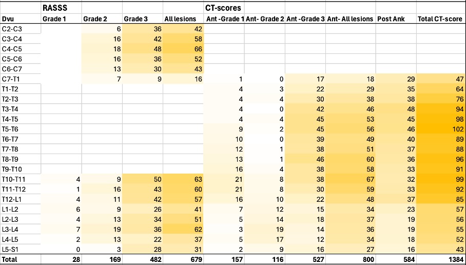
monocentric restrospective study included AS patients responding to modified New York criteria for radiographic sacroiliitis. Patients were required to have undergone spine x-ray and a TAP CT within a delay of 12 months. On TAP CT, the 36 anterior vertebra corner from C7 to S1 were graded for erosion/squaring (grade 1), syndesmophytes (grade 2) and bone bridge (grade 3). The anterior CT score ranged from 0 to 108 (sum of the grades). The posterior segment evaluated facet ankylosis as present (grade 3) or absent (grade 0). The posterior CT score ranged from 0 to 108 (sum of the grades). The total CT-score (ant+post) ranged from 0 to 216.
Spine radiographic structural assessment was established by one experienced reader on sagittal x-rays according to the RASSS (from 0-84).

Results: 142 patients were included: median age 54 y [47-62], 70.4% male, 66.6% smokers, 77.6%  HLA-B27 status, median duration disease 14 y [6-26], 43.5% treated by bDMARDS. Among them, 90 (62.7%) and 102 (71.8%) patients presented spine structural damage according to the RASSS and the total CT-score respectively (p=0.0001). For the total CT-score, 94 patients presented at least one lesion at the anterior vertebra corner and 68 with facet joint ankylosis. The sensitivity, the specificity and the accuracy of the CT score were 60.0%, 72.5% and 69% respectively with RASSS as referent score.  The mean RASSS and total CT-scores were 13.1 (± 20.0) and the 37.2 ((± 47.2) respectively. The correlation between RASSS and total CT score was excellent (value= 0.83; p< 0.0001). For RASSS, linear regression analysis showed associations with age (value =0.23; p=0.01), disease duration (value 0.27; p< 0.002), male gender (value =8.4 p=0.01) and CRP >5 mg/l (value = 0.17; p=0.03). The total-CT score showed association with age (value=0.17; p=0.034), male gender (value=0.16; p=0.03) disease duration (value=0.31; p=0.0001), and CRP >5 mg/l (value=0.25; p=0.001) in multivariate analysis. Reading was performed with excellent intra reader reliabilities for both radiographic and CT assessment (ICCs >0.90).

Conclusion: In AS patients, screening spine structural lesion on opportunistic TAP CT is an efficient, reproductible and complementary approach to x-ray imaging. In our study, CT screening allowed anterior and posterior dorsolombar spine assessment, which can be challenging to achieve with x-rays, with a greater number of spine lesions to be detected.

Supporting image 1

Figure 1: Heat map of spine structural lesions prevalence and severity at discovertebral unit (DVu) level on radiography (RASSS: Radiographic Ankylosing Spondylitis Spinal Score) and on CT. On CT, the anterior segment was graded on the presence of erosion/squaring (Ant-Grade 1), syndesmophytes (Ant-Grade 2) and/or spinal ankylosis with bone bridging (Ant-Grade 3) for each vertebra. The anterior score (Ant-All lesions) varied from 0 to 108. The posterior segments evaluated facet ankylosis according to a binary manner for each DVu (present: 3 points; or absent: 0 point). The posterior score (Post Ank) varied also from 0 to 108. The total CT-score was the sum of the anterior and posterior scores and ranged from 0 to 216.


Disclosures: C. Morizot: None; I. Favre-Felix: None; A. Mennini: None; E. Bauer: None; I. Chary-Valckenaere: None; D. Loeuille: None.

To cite this abstract in AMA style:

Morizot C, Favre-Felix I, Mennini A, Bauer E, Chary-Valckenaere I, Loeuille D. Spine Structural Lesions in Ankylosing Spondylitis: Comparison Between Radiographic (RASSS) and Opportunistic Thoraco-abdomino-pelvic CT Assessment [abstract]. Arthritis Rheumatol. 2024; 76 (suppl 9). https://acrabstracts.org/abstract/spine-structural-lesions-in-ankylosing-spondylitis-comparison-between-radiographic-rasss-and-opportunistic-thoraco-abdomino-pelvic-ct-assessment/. Accessed .
  • Tweet
  • Click to email a link to a friend (Opens in new window) Email
  • Click to print (Opens in new window) Print

« Back to ACR Convergence 2024

ACR Meeting Abstracts - https://acrabstracts.org/abstract/spine-structural-lesions-in-ankylosing-spondylitis-comparison-between-radiographic-rasss-and-opportunistic-thoraco-abdomino-pelvic-ct-assessment/

Advanced Search

Your Favorites

You can save and print a list of your favorite abstracts during your browser session by clicking the “Favorite” button at the bottom of any abstract. View your favorites »

All abstracts accepted to ACR Convergence are under media embargo once the ACR has notified presenters of their abstract’s acceptance. They may be presented at other meetings or published as manuscripts after this time but should not be discussed in non-scholarly venues or outlets. The following embargo policies are strictly enforced by the ACR.

Accepted abstracts are made available to the public online in advance of the meeting and are published in a special online supplement of our scientific journal, Arthritis & Rheumatology. Information contained in those abstracts may not be released until the abstracts appear online. In an exception to the media embargo, academic institutions, private organizations, and companies with products whose value may be influenced by information contained in an abstract may issue a press release to coincide with the availability of an ACR abstract on the ACR website. However, the ACR continues to require that information that goes beyond that contained in the abstract (e.g., discussion of the abstract done as part of editorial news coverage) is under media embargo until 10:00 AM ET on November 14, 2024. Journalists with access to embargoed information cannot release articles or editorial news coverage before this time. Editorial news coverage is considered original articles/videos developed by employed journalists to report facts, commentary, and subject matter expert quotes in a narrative form using a variety of sources (e.g., research, announcements, press releases, events, etc.).

Violation of this policy may result in the abstract being withdrawn from the meeting and other measures deemed appropriate. Authors are responsible for notifying colleagues, institutions, communications firms, and all other stakeholders related to the development or promotion of the abstract about this policy. If you have questions about the ACR abstract embargo policy, please contact ACR abstracts staff at [email protected].

Wiley

  • Online Journal
  • Privacy Policy
  • Permissions Policies
  • Cookie Preferences

© Copyright 2025 American College of Rheumatology