Session Information
Session Type: Poster Session A
Session Time: 9:00AM-11:00AM
Background/Purpose: Magnetic resonance imaging (MRI) is frequently used in patients with chronic back pain (CBP) to diagnose mechanical and inflammatory diseases, including axial spondyloarthritis (axSpA). Recent data suggest sacroiliac joints (SIJ) MRI findings in axSpA may differ according to sex1. Whether this holds true for spinal MRI and other causes of CBP is currently unknown. Our aim was to analyze spinal and SIJ MRI findings in young male and female patients with CBP.
Methods: The “Strategy for a Hospital Early Referral in Patients with Axial Spondyloarthritis” (SHERPAS) is a prospective ongoing study recruiting young patients (18 to 40 years) with CBP asked to undergo an MRI of the spine by other specialists different than rheumatologists in a tertiary hospital, starting in September 2021. After inclusion, an additional MRI of the SIJ, followed by a rheumatology visit and eligible blood tests were performed. Dataset for this interim analysis was locked in October 2022. The protocol for MRI of the spine used a 1.5T scanner to acquire sagittal T1-weighted turbo spin echo (TSE) and T2- weighted TSE, both for the lateral sides of vertebral bodies, and T2-Multistack. On top of this, an MRI of the SIJ, which involved T1-TSE, T2- weighted SPAIR and short-tau inversion recovery (STIR) sequences, was performed. MRI findings were assessed according to clinical practice by one of the four musculoskeletal radiologists working in the centre, describing the presence of mechanical lesions (spondylolisthesis, spondylolysis, disc herniation, disc protrusion, facet joints abnormalities), inflammatory findings (both SIJ and spine), and SIJ structural findings. For this analysis, results were stratified by sex.
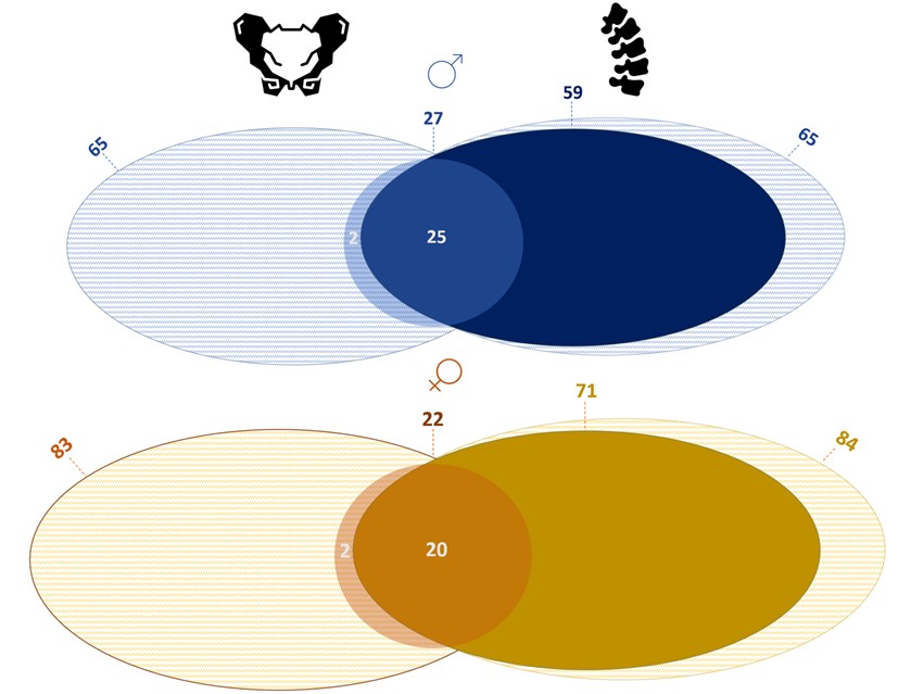
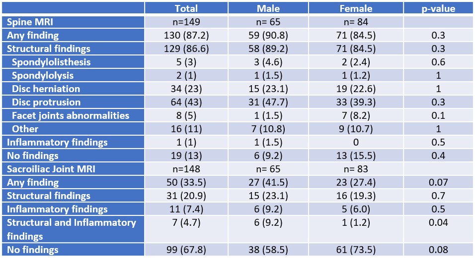
Results: Among152 recruited patients, 85 (55.9%) were female; mean age was 34.2 (5.3) years. Spinal MRI findings were reported in 130 (87.2%) patients, with no differences between sexes (male 90.8% vs female 84.5%, p=0.3). As shown in Table, the most frequent diagnosis in both sexes were disc protrusion, followed by disc herniation. Inflammatory spinal findings were detected only in one male patient. No differences were found for any of the spinal lesions. SIJ MRI findings were reported in 49 (33.1%) patients, being numerically more frequently observed in males than females (41.5% vs 26.5%; p=0.08). Concurrence of both structural and inflammatory lesions was more frequently in males (9.2% vs 1.2%, p< 0.05), while no differences in isolate SIJ MRI findings were found between sexes. Overall, 45 (29.6%) patients presented findings both in the spine and SIJ (Figure).
Conclusion: In young patients who are requested a spinal MRI by other specialists different than rheumatologists, spinal lesions are reported in most patients, and similarly in males and females. Remarkably, SIJ MRI findings are reported in one out of three patients in this population, being more frequently in males.
To cite this abstract in AMA style:
Benavent D, Tapia M, muley V, Bernabeu D, Plasencia-Rodríguez C, Balsa A, Navarro-Compán V. Spine and Sacroiliac Joints Findings in Young Males and Females with Chronic Back Pain Undergoing Magnetic Resonance Imaging in Clinical Practice [abstract]. Arthritis Rheumatol. 2023; 75 (suppl 9). https://acrabstracts.org/abstract/spine-and-sacroiliac-joints-findings-in-young-males-and-females-with-chronic-back-pain-undergoing-magnetic-resonance-imaging-in-clinical-practice/. Accessed .« Back to ACR Convergence 2023
ACR Meeting Abstracts - https://acrabstracts.org/abstract/spine-and-sacroiliac-joints-findings-in-young-males-and-females-with-chronic-back-pain-undergoing-magnetic-resonance-imaging-in-clinical-practice/


