ACR Meeting Abstracts

ACR Meeting Abstracts

  • Meetings
    • ACR Convergence 2024
    • ACR Convergence 2023
    • 2023 ACR/ARP PRSYM
    • ACR Convergence 2022
    • ACR Convergence 2021
    • ACR Convergence 2020
    • 2020 ACR/ARP PRSYM
    • 2019 ACR/ARP Annual Meeting
    • 2018-2009 Meetings
    • Download Abstracts
  • Keyword Index
  • Advanced Search
  • Your Favorites
    • Favorites
    • Login
    • View and print all favorites
    • Clear all your favorites
  • ACR Meetings

Abstract Number: 1150

Spatial Transcriptomics Stratifies Health and Psoriatic Disease Severity by Emergent Cellular Ecosystems

Rochelle Castillo1, Ikjot Sidhu1, Igor Dolgalev1, Ipsita Subudhi1, Di Yan1, Piotr Konieczny1, Brandon Hsieh1, Tinyi Chu2, Rebecca Haberman1, Shanmugapriya Selvaraj1, Tomoe Shiomi1, Rhina Medina1, Parvathy Vasudevanpillai Girija1, Adriana Heguy1, Cynthia Loomis1, Luis Chiriboga1, Shane Meehan1, Christopher Ritchlin3, Maria de la Luz Garcia-Hernandez4, John Carucci1, Andrea Neimann1, Shruti Naik1 and Jose Scher5, 1NYU Langone Health, New York, NY, 2Memorial Sloan Kettering Cancer Center, New York, NY, 3Allergy, Immunology and Rheumatology Division, University of Rochester Medical School, Canandaigua, NY, 4University of Rochester, West Henrietta, NY, 5New York University School of Medicine, New York, NY

Meeting: ACR Convergence 2022

Keywords: Bioinformatics, Dermatology, Gene Expression, Psoriatic arthritis, skin

  • Tweet
  • Click to email a link to a friend (Opens in new window) Email
  • Click to print (Opens in new window) Print
Session Information

Date: Sunday, November 13, 2022

Title: Spondyloarthritis Including PsA – Basic Science Poster

Session Type: Poster Session C

Session Time: 1:00PM-3:00PM

Background/Purpose: The skin is recognized as a window into the immunopathogenic mechanisms driving the vast phenotypic spectrum of psoriatic disease.

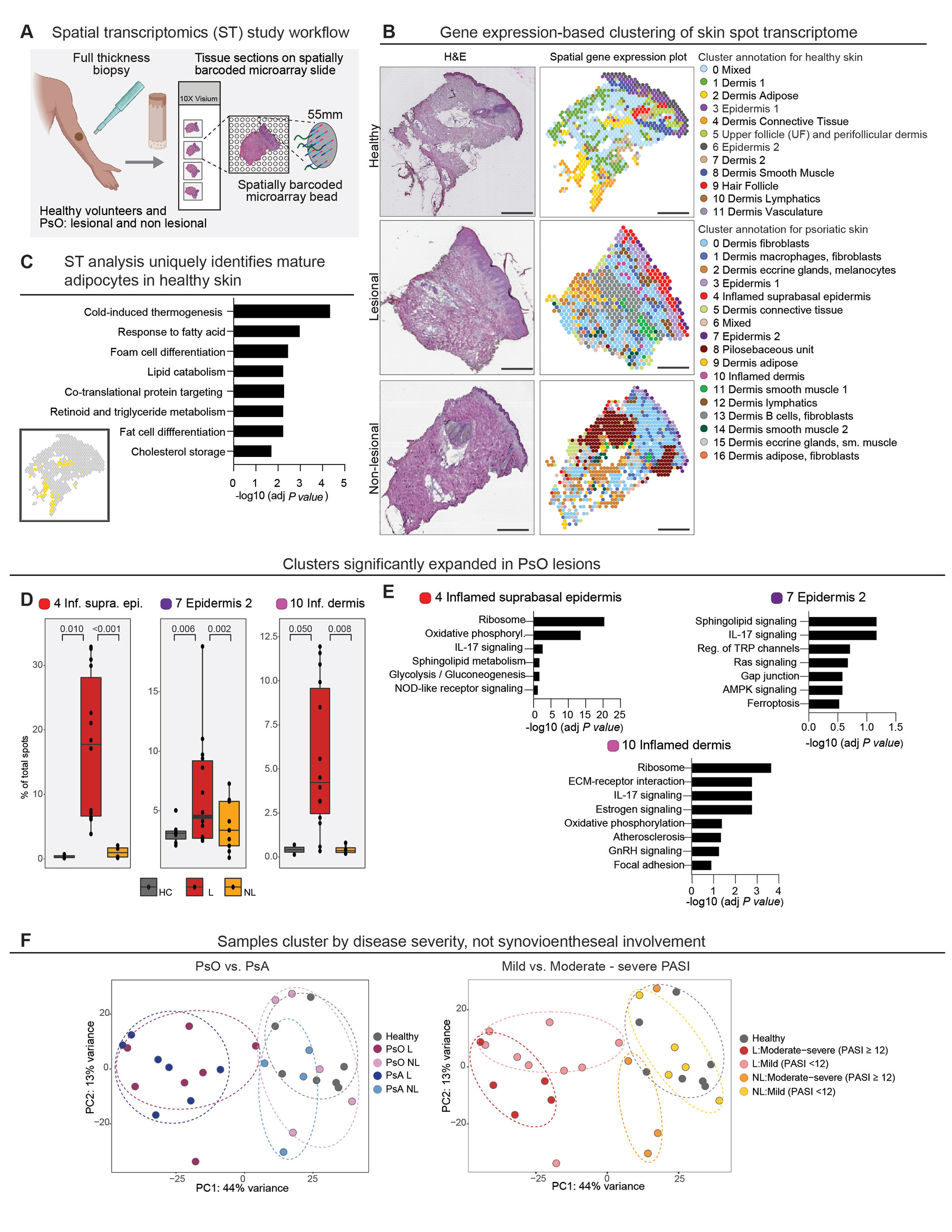
Methods: To better decipher the cellular landscape of both healthy and psoriatic skin, we employed spatial transcriptomics (ST), a ground-breaking technology that precisely maps gene expression from histologically-intact tissue sections (Fig. 1A).

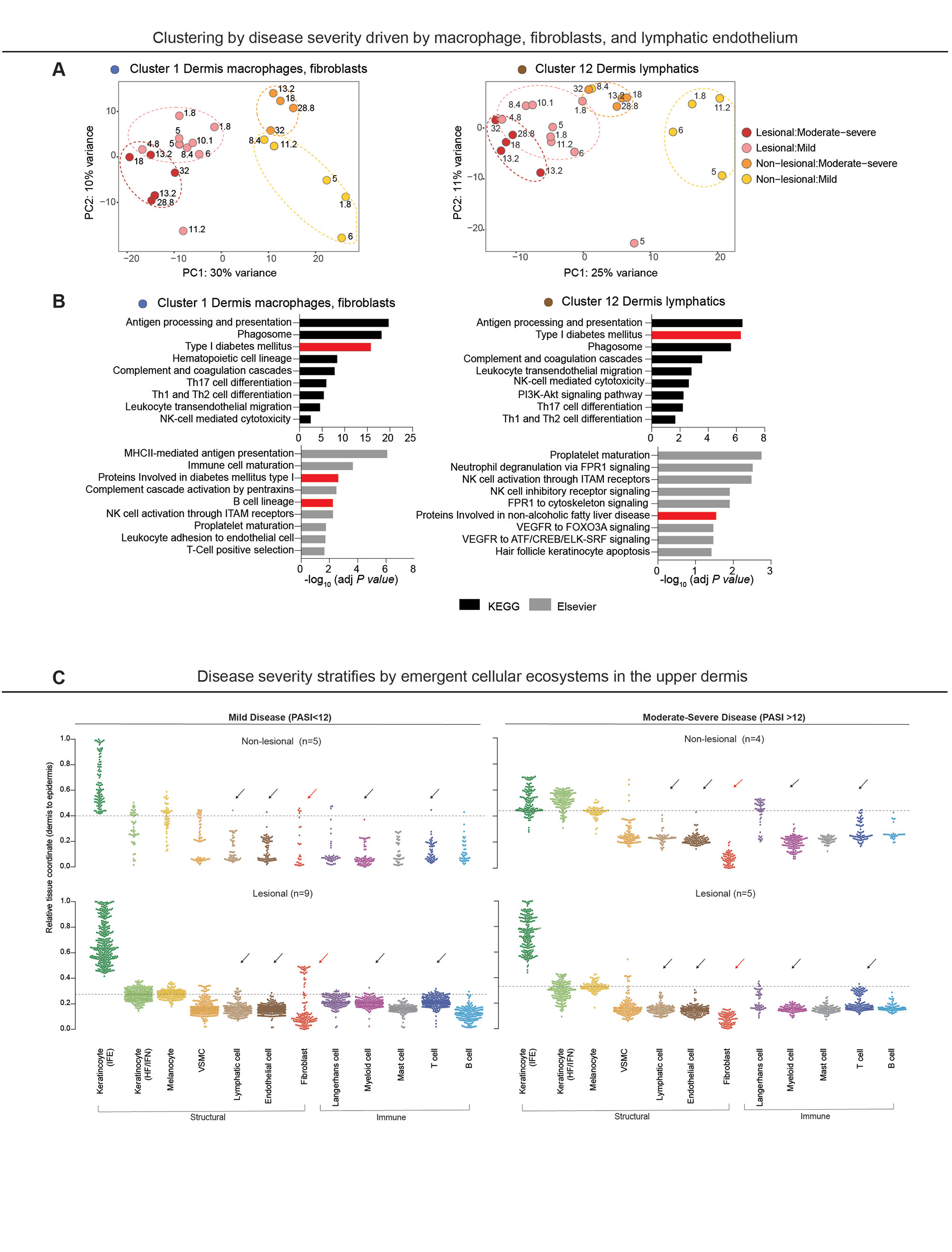
Results: Findings gleaned from computationally integrating our 23 matched lesional and non-lesional psoriatic and 7 healthy control samples with publicly-available single-cell ribonucleic acid (RNA) sequencing datasets established the ability of ST to recapitulate the tissue architecture of both healthy and inflamed skin (Fig. 1B) and highlighted topographic shifts in the immune cell milieu, from a predominantly perifollicular distribution in steady-state skin to the papillary and upper reticular dermis in psoriatic lesional skin. We also incidentally discovered that ST’s ability to ascertain gene expression patterns from intact tissue rendered it particularly conducive to studying the transcriptome of lipid-laden cells such as dermal adipose tissue and sebaceous glands (Fig. 1C), whose expression profiles are typically lost in the process of tissue handling and dissociation for bulk and single-cell RNA seq. Unbiased clustering of pooled healthy and psoriatic samples identified two epidermal clusters and one dermal cluster that were differentially expanded in psoriatic lesional skin (p values ≤0.05) (Fig. 1D); pathway analysis of these clusters revealed enrichment of known psoriatic inflammatory pathways (Fig. 1E). Unsupervised classification of skin-limited psoriasis and psoriatic arthritis samples revealed stratification by cutaneous disease severity or Psoriasis Area and Severity Index (PASI) score and not by presence or absence of concomitant systemic/synovial disease (Fig. 1F). Remarkably, this PASI-dependent segregation was also evident in distal, non-lesional samples and was driven by the dermal macrophage and fibroblast cluster and the lymphatic endothelium (Fig. 2A). Inquiry into the mechanistic drivers of this observed stratification yielded enrichment of pathways associated with key T cell and innate immune cell activation, B cells, and metabolic dysfunction (Fig. 2B). Finally, tissue scale computational cartography of gene expression revealed differences in regional enrichment of specific cell types across phenotypic groups, most notably upward extension of fibroblasts to the upper dermis in both lesional and non-lesional samples from mild psoriasis and restriction to the lower dermis in the moderate-to-severe psoriasis samples (Fig. 2C), suggesting that disease severity stratification may be driven by emergent cellular ecosystems in the upper dermis.

Conclusion: Thus, we have been able to successfully leverage ST integrated with independently-generated single-cell RNA seq data to spatially define the emergent cellular ecosystems of healthy and matched psoriatic lesional and non-lesional skin and in so doing, demonstrated the value of ST in unearthing the genetic groundwork at both the site of inflammation and in distal, clinically-uninvolved skin.

Supporting image 1

Fig. 1. (A) Schematic of spatial transcriptomics study workflow. Four mm skin punch biopsies were obtained from healthy volunteers (n=3) and lesional and non-lesional skin from patients with psoriatic disease (n=11). Ten micron-thick sections were then placed on capture areas on the ST microarray slide, each containing molecularly barcoded, spatially encoded spots with a diameter of 50 microns and a center-to-center distance of 100 microns. (B) Side-by-side comparison of a hematoxylin-eosin (H&E) stained section of representative healthy, lesional, and non-lesional skin samples and the corresponding ST plots showed concordance of unbiased gene expression-based clustering with histologic tissue architecture. (C) Pathway analysis of the adipose cluster in healthy skin (cluster 2) confirmed upregulation of lipid-associated processes. Inset: Spots corresponding to the adipose cluster highlighted in yellow. (D) Wilcoxon rank sum test (results displayed as box plots) yielded statistically significant expansion of three clusters in lesional skin compared to both non-lesional and healthy skin – inflamed suprabasal epidermis (cluster 4), epidermis 2 (cluster 7), and inflamed dermis (cluster 10). HC=healthy control, L=lesional psoriatic skin, NL=non-lesional psoriatic skin. (E) Pathways enriched in clusters 4, 7, and 10. (F) Principal component analysis (PCA) plots demonstrating segregation of samples by severity of cutaneous disease in both lesional and non-lesional samples along the first principal component (right) that was not seen in the samples categorized according to presence or absence of arthritis (left). PsA=psoriatic arthritis, PsO=skin-limited psoriasis.

Supporting image 2

Fig. 2. (A) PCA of lesional and non-lesional samples colored by disease severity in spatial clusters 1 (left) and 12 (right) revealed more discrete clustering. (B) Pathways significantly enriched in clusters 1 (left) and 12 (right) showed enrichment of pathways associated with key T cell and innate immune cell activation, B cells, and metabolic dysfunction (highlighted in red). (C) SpaceFold one dimension projection of cell distribution from an independently-generated single-cell RNA seq data set on aggregated ST lesional and non-lesional samples from mild (PASI-low) and moderate-severe (PASI-high) samples. Y-axis represents tissue position, starting with the lower dermis marked as position 0 to suprabasal epidermis marked as position 1. Dashed line represents epidermal-dermal junction, discerned by cell types in the basal epidermal layer (melanocytes and Langerhans cells). Fibroblast signatures (red arrows) were largely relegated to the lower dermis in the PASI-high group, but extended to the upper dermis in the PASI-low group. This striking difference in fibroblast localization was also noted in non-lesional PASI-high vs. PASI-low groups. In addition to fibroblasts, lymphatic, endothelial, myeloid, and T cells signatures (black arrows) were also observed in the upper dermis of lesional PASI-low samples, but were much lower in the dermis of PASI-low non-lesional and all samples in the PASI-high group. Interfollicular epidermis (IFE), hair follicle and infundibulum (HF/IFN), n= number of individual biopsies.


Disclosures: R. Castillo, None; I. Sidhu, None; I. Dolgalev, None; I. Subudhi, None; D. Yan, None; P. Konieczny, None; B. Hsieh, None; T. Chu, None; R. Haberman, None; S. Selvaraj, None; T. Shiomi, None; R. Medina, None; P. Vasudevanpillai Girija, None; A. Heguy, None; C. Loomis, None; L. Chiriboga, None; S. Meehan, None; C. Ritchlin, UCB, AbbVie, Eli Lilly, Pfizer Inc, Novartis, Janssen, Bristol-Myers Squibb; M. Garcia-Hernandez, None; J. Carucci, Genentech; A. Neimann, Janssen, UCB, AbbVie/Abbott, Bristol-Myers Squibb(BMS); S. Naik, None; J. Scher, Janssen, Pfizer, AbbVie, Sanofi, Novartis, UCB.

To cite this abstract in AMA style:

Castillo R, Sidhu I, Dolgalev I, Subudhi I, Yan D, Konieczny P, Hsieh B, Chu T, Haberman R, Selvaraj S, Shiomi T, Medina R, Vasudevanpillai Girija P, Heguy A, Loomis C, Chiriboga L, Meehan S, Ritchlin C, Garcia-Hernandez M, Carucci J, Neimann A, Naik S, Scher J. Spatial Transcriptomics Stratifies Health and Psoriatic Disease Severity by Emergent Cellular Ecosystems [abstract]. Arthritis Rheumatol. 2022; 74 (suppl 9). https://acrabstracts.org/abstract/spatial-transcriptomics-stratifies-health-and-psoriatic-disease-severity-by-emergent-cellular-ecosystems/. Accessed .
  • Tweet
  • Click to email a link to a friend (Opens in new window) Email
  • Click to print (Opens in new window) Print

« Back to ACR Convergence 2022

ACR Meeting Abstracts - https://acrabstracts.org/abstract/spatial-transcriptomics-stratifies-health-and-psoriatic-disease-severity-by-emergent-cellular-ecosystems/

Advanced Search

Your Favorites

You can save and print a list of your favorite abstracts during your browser session by clicking the “Favorite” button at the bottom of any abstract. View your favorites »

All abstracts accepted to ACR Convergence are under media embargo once the ACR has notified presenters of their abstract’s acceptance. They may be presented at other meetings or published as manuscripts after this time but should not be discussed in non-scholarly venues or outlets. The following embargo policies are strictly enforced by the ACR.

Accepted abstracts are made available to the public online in advance of the meeting and are published in a special online supplement of our scientific journal, Arthritis & Rheumatology. Information contained in those abstracts may not be released until the abstracts appear online. In an exception to the media embargo, academic institutions, private organizations, and companies with products whose value may be influenced by information contained in an abstract may issue a press release to coincide with the availability of an ACR abstract on the ACR website. However, the ACR continues to require that information that goes beyond that contained in the abstract (e.g., discussion of the abstract done as part of editorial news coverage) is under media embargo until 10:00 AM ET on November 14, 2024. Journalists with access to embargoed information cannot release articles or editorial news coverage before this time. Editorial news coverage is considered original articles/videos developed by employed journalists to report facts, commentary, and subject matter expert quotes in a narrative form using a variety of sources (e.g., research, announcements, press releases, events, etc.).

Violation of this policy may result in the abstract being withdrawn from the meeting and other measures deemed appropriate. Authors are responsible for notifying colleagues, institutions, communications firms, and all other stakeholders related to the development or promotion of the abstract about this policy. If you have questions about the ACR abstract embargo policy, please contact ACR abstracts staff at [email protected].

Wiley

  • Online Journal
  • Privacy Policy
  • Permissions Policies
  • Cookie Preferences

© Copyright 2025 American College of Rheumatology