ACR Meeting Abstracts

ACR Meeting Abstracts

  • Meetings
    • ACR Convergence 2024
    • ACR Convergence 2023
    • 2023 ACR/ARP PRSYM
    • ACR Convergence 2022
    • ACR Convergence 2021
    • ACR Convergence 2020
    • 2020 ACR/ARP PRSYM
    • 2019 ACR/ARP Annual Meeting
    • 2018-2009 Meetings
    • Download Abstracts
  • Keyword Index
  • Advanced Search
  • Your Favorites
    • Favorites
    • Login
    • View and print all favorites
    • Clear all your favorites
  • ACR Meetings

Abstract Number: 0642

SLAMF6-SAP Signaling Unit Is Increased in SLE T Follicular Helper Cells

Yevgeniya Gartshteyn1, Leila Khalili2, Adam Mor3 and Anca Askanase2, 1Columbia University Medical Center, Glen Rock, NJ, 2Columbia University Medical Center, New York, NY, 3Columbia University, New York, NY

Meeting: ACR Convergence 2022

Keywords: signal transduction, Systemic lupus erythematosus (SLE), T Cell

  • Tweet
  • Click to email a link to a friend (Opens in new window) Email
  • Click to print (Opens in new window) Print
Session Information

Date: Sunday, November 13, 2022

Title: SLE – Etiology and Pathogenesis Poster

Session Type: Poster Session B

Session Time: 9:00AM-10:30AM

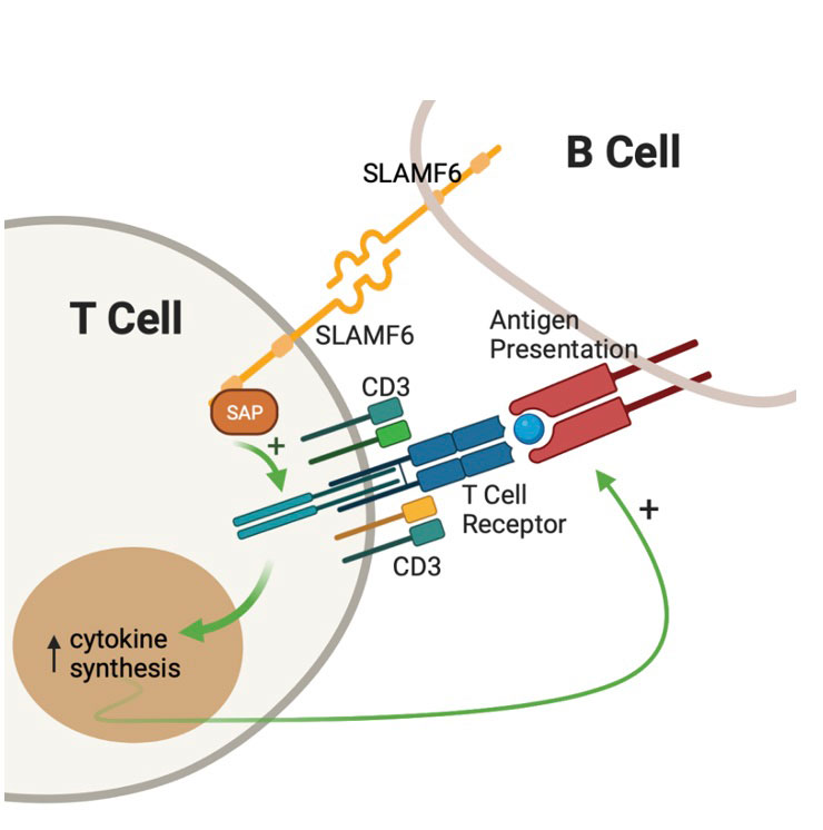
Background/Purpose: SLE is a multiorgan disease in which immune cells lack self-tolerance resulting in autoimmunity. SLE T cells infiltrate organs, provide help to autoreactive B cells and mediate inflammatory response. Signaling Lymphocyte Activation Molecule 6 (SLAMF6) is a T cell co-receptor whose gene is a risk locus for SLE both in murine models and humans. Following SLAMF6 receptor ligation, the intracellular signal is transmitted and amplified by the SLAM Associated Protein (SAP) –Fig1. SLAMF6-SAP signaling is critical for healthy T-B cell interactions, germinal center formations, and B cell maturation. This study was initiated to evaluate the role of SLAMF6-SAP in SLE.

Methods: Peripheral blood mononuclear cells (PBMCs) were isolated from whole blood using density gradient separation. Immunophenotyping using flow cytometry was performed after cells were labeled with anti-CD3 (FITC), anti-CD4 (AF700), anti-CD8 (BV605), anti-CXCR5 (PE-Cy7), anti-PD1 (BV421) and either anti-SLAMF6 or anti-SAP (APC). PBMCs were activated with anti-CD3 ± anti-SLAMF6 for 24 hours, after which IL-2 levels were analyzed by ELISA. RNA sequencing data from SLE patients was accessed from Gene Expression Omnibus (GEO) and analyzed for SAP gene (SH2D1A) expression.

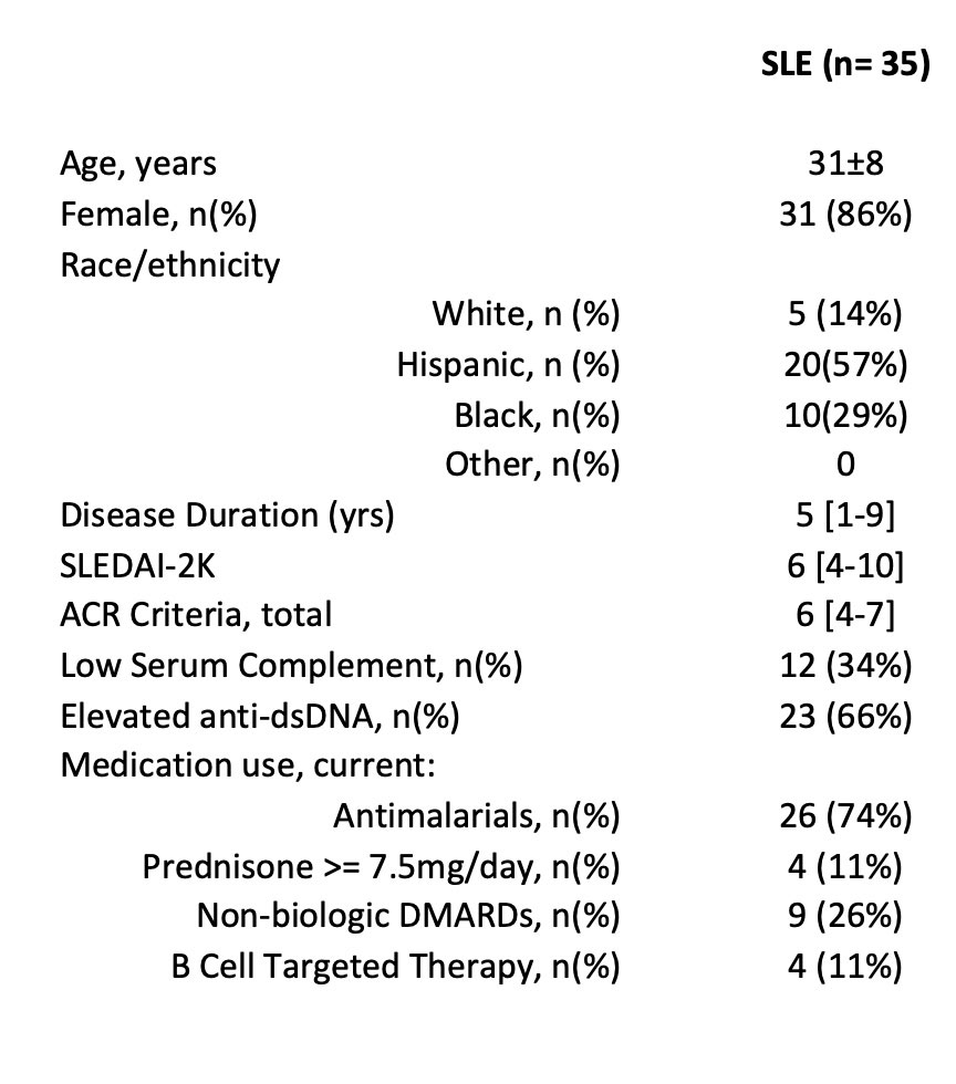
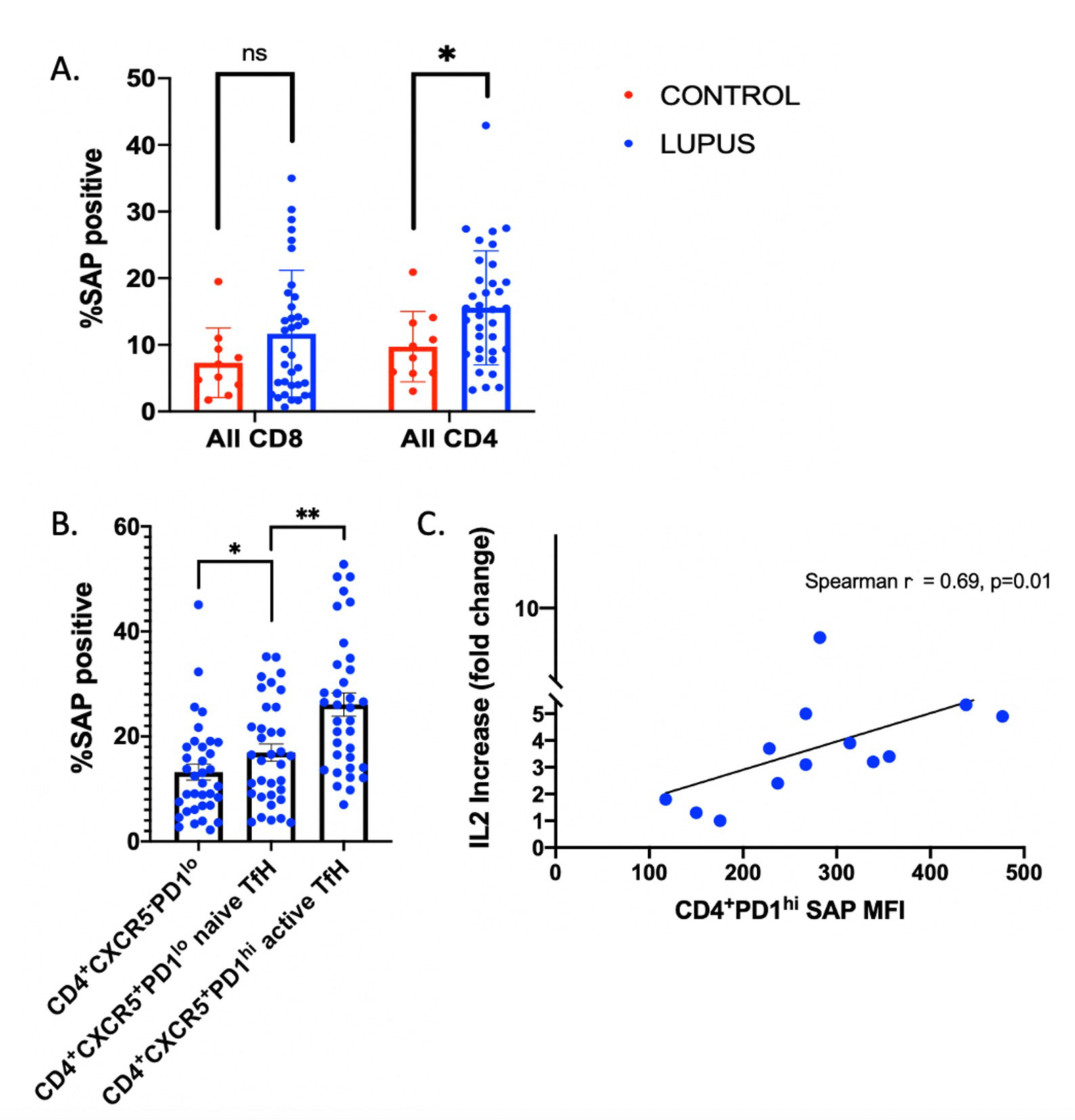
Results: We enrolled 35 patients with SLE (1997 ACR criteria), ages 31±8, 86% female. Median SLE disease duration was 5 [1-9] years. Median SLEDAI was 6 [4-10]; 34% had low complements and 66% had elevated anti-dsDNA. 74% were taking antimalarials, 11% prednisone 7.5mg/day, 26% non-biologic DMARDs, and 11% B-cell targeted therapy. Detailed data is presented in Table 1. SLAMF6 was not differentially expressed between SLE and healthy control CD4 T cells (93.7% and 93.2%, respectively, p=0.9). Conversely, SAP expression was increased in SLE CD4, but not CD8, T cells as compared to healthy controls (15.6% vs 9.8% p< 0.05 and 11.7% vs 7.3% p=0.2, respectively) -Fig 2A. SAP levels were increased in CD4+CXCR5+ T-follicular helper cells (TfH) as compared to CD4+CXCR5– cells (p=0.03), with still higher SAP levels seen in the activated TfH subgroup, defined by PD-1 expression (26.1% of CD4+CXCR5+PD1hi vs. 13.2% of CD4+CXCR5+PD1lo cells, p< 0.001) -Fig 2B. Stimulation of PBMCs with anti-CD3 + anti-SLAMF6 vs. anti-CD3 alone resulted in fold change IL-2 increase that correlated with CD4+PD1hi SAP expression (p=0.01) -Fig 2C. RNA sequencing data identified increased SAP expression in SLE as compared to control groups.

Conclusion: SLE T cells express greater SAP levels that result in enhanced SLAMF6 signaling. This is especially pronounced in the TfH cells, highlighting the role of TfH in SLE pathogenesis and identifying SLAMF6 as a potentially novel therapeutic target in the future.

Supporting image 1

Table 1. Clinical and demographic characteristics.

Supporting image 2

Figure 1. SLAMF6 co-receptor modulates T cell receptor signaling. SLAMF6 ligation results in recruitment of SLAM Associated Protein (SAP) leading to downstream enhancement of T Cell Receptor activation, cytokine release, and stabilized T-B cell cross-talk.

Supporting image 3

Figure 2. SAP levels are increased in activated SLE T-follicular helper cells (TfH). A) Peripheral blood mononuclear cells were labeled to identify the CD3+CD4+CD8- and CD3+CD4-CD8+ T-Cell populations using flow cytometry. The frequency of SAP positive cells is reported. B) CXCR5+ TfH were identified by flow cytometry. PD1-high (PD1hi) expression identified the activated, or antigen-primed, TfH cell subpopulation. The frequency of SAP positive cells is reported. C) Peripheral blood mononuclear cells (PBMCs) from SLE patients were stimulated with anti-CD3 + IgG-ISO (control) vs. anti-CD3+anti-SLAMF6 antibodies. Interleukin_2 (IL_2) levels were measured by ELISA. The fold change increase in interleukin 2 (IL_2) release in the presence of anti-SLAMF6 activation vs. anti-CD3 alone is shown.


Disclosures: Y. Gartshteyn, None; L. Khalili, None; A. Mor, None; A. Askanase, AstraZeneca, GlaxoSmithKlein(GSK), Aurinia, Amgen, Pfizer, Idorsia, Eli Lilly, UCB, AbbVie/Abbott, Janssen, Bristol-Myers Squibb(BMS).

To cite this abstract in AMA style:

Gartshteyn Y, Khalili L, Mor A, Askanase A. SLAMF6-SAP Signaling Unit Is Increased in SLE T Follicular Helper Cells [abstract]. Arthritis Rheumatol. 2022; 74 (suppl 9). https://acrabstracts.org/abstract/slamf6-sap-signaling-unit-is-increased-in-sle-t-follicular-helper-cells/. Accessed .
  • Tweet
  • Click to email a link to a friend (Opens in new window) Email
  • Click to print (Opens in new window) Print

« Back to ACR Convergence 2022

ACR Meeting Abstracts - https://acrabstracts.org/abstract/slamf6-sap-signaling-unit-is-increased-in-sle-t-follicular-helper-cells/

Advanced Search

Your Favorites

You can save and print a list of your favorite abstracts during your browser session by clicking the “Favorite” button at the bottom of any abstract. View your favorites »

All abstracts accepted to ACR Convergence are under media embargo once the ACR has notified presenters of their abstract’s acceptance. They may be presented at other meetings or published as manuscripts after this time but should not be discussed in non-scholarly venues or outlets. The following embargo policies are strictly enforced by the ACR.

Accepted abstracts are made available to the public online in advance of the meeting and are published in a special online supplement of our scientific journal, Arthritis & Rheumatology. Information contained in those abstracts may not be released until the abstracts appear online. In an exception to the media embargo, academic institutions, private organizations, and companies with products whose value may be influenced by information contained in an abstract may issue a press release to coincide with the availability of an ACR abstract on the ACR website. However, the ACR continues to require that information that goes beyond that contained in the abstract (e.g., discussion of the abstract done as part of editorial news coverage) is under media embargo until 10:00 AM ET on November 14, 2024. Journalists with access to embargoed information cannot release articles or editorial news coverage before this time. Editorial news coverage is considered original articles/videos developed by employed journalists to report facts, commentary, and subject matter expert quotes in a narrative form using a variety of sources (e.g., research, announcements, press releases, events, etc.).

Violation of this policy may result in the abstract being withdrawn from the meeting and other measures deemed appropriate. Authors are responsible for notifying colleagues, institutions, communications firms, and all other stakeholders related to the development or promotion of the abstract about this policy. If you have questions about the ACR abstract embargo policy, please contact ACR abstracts staff at [email protected].

Wiley

  • Online Journal
  • Privacy Policy
  • Permissions Policies
  • Cookie Preferences

© Copyright 2025 American College of Rheumatology