ACR Meeting Abstracts

ACR Meeting Abstracts

  • Meetings
    • ACR Convergence 2024
    • ACR Convergence 2023
    • 2023 ACR/ARP PRSYM
    • ACR Convergence 2022
    • ACR Convergence 2021
    • ACR Convergence 2020
    • 2020 ACR/ARP PRSYM
    • 2019 ACR/ARP Annual Meeting
    • 2018-2009 Meetings
    • Download Abstracts
  • Keyword Index
  • Advanced Search
  • Your Favorites
    • Favorites
    • Login
    • View and print all favorites
    • Clear all your favorites
  • ACR Meetings

Abstract Number: 2373

SIRPα Expression in Systemic Vasculitis

Shubhasree Banerjee1, Eileen Rose2, Sandip Panicker2, Nader Khalidi3, Curry Koening4, Carol Langford5, Paul Monach6, Christian Pagnoux7, Carol McAlear1 and Peter Merkel1, 1University of Pennsylvania, Philadelphia, PA, 2Electra Therapeutics, Inc., South San Francisco, CA, 3McMaster University, Hamilton, ON, Canada, 4University of Texas Dell Medical School, Austin, TX, 5Cleveland Clinic, Cleveland, OH, 6VA Boston Healthcare System, Boston, MA, 7Mount Sinai Hospital, Toronto, ON, Canada

Meeting: ACR Convergence 2023

Keywords: ANCA associated vasculitis, giant cell arteritis, Vasculitis

  • Tweet
  • Click to email a link to a friend (Opens in new window) Email
  • Click to print (Opens in new window) Print
Session Information

Date: Tuesday, November 14, 2023

Title: (2370–2386) Vasculitis – ANCA-Associated Poster III: Biomarkers & Renal Outcomes

Session Type: Poster Session C

Session Time: 9:00AM-11:00AM

Background/Purpose: Signal regulatory protein alpha (SIRPα) is primarily found on myeloid cells, including macrophages and neutrophils. Upon binding to CD47, SIRPa signaling regulates various cellular functions, including phagocytosis, antigen presentation, cell proliferation, cellular fusion, and cell migration. Therefore, SIRPamay be involved in the pathogenesis of autoimmune diseases including systemic vasculitis. This study aimed to assess SIRPa expression in tissue samples from patients with vasculitis.

Methods: Immunohistochemical staining for SIRPα was performed on kidney, lung, and temporal artery (TA) biopsy samples from patients with granulomatosis with polyangiitis (GPA), microscopic polyangiitis (MPA), giant cell arteritis (GCA), and patients without vasculitis. Cells were grouped into 4 categories: i) monocytes/macrophages/dendritic cells; ii) neutrophils; iii) eosinophils; and iv) lymphocytes. Categories iii and iv were not analyzed because of positive isotype control staining and absence of SIRPα+ cells, respectively. A semiquantitative estimation of SIRPα+ cells in categories i and ii were reported using the H-score system based on a visual estimate of the percentage of SIRPα+ cells and the staining intensities classified from 0 to 3 (H-score = 0 x [% with 0 intensity] + 1 x [% with 1 intensity] + 2 x [% with 2 intensity] + 3 x [% with 3 intensity]). Density of inflammatory infiltrate for individual cell categories were visually estimated and reported using an ordinal scale. Statistical analysis was done using GraphPad Prism 9.5.

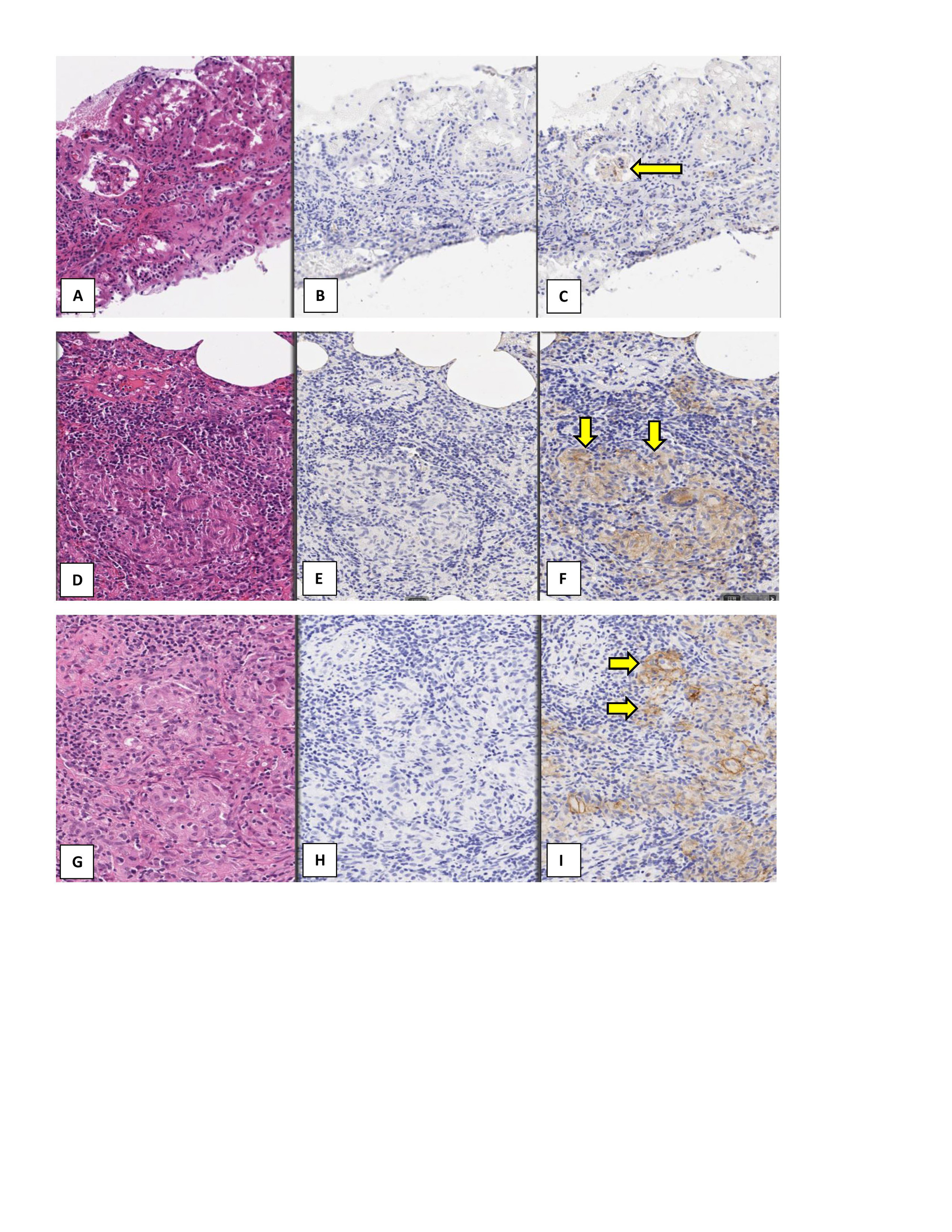
Results: Samples from 43 patients (20 with GPA, 8 with MPA, and 15 with GCA) were included in the study. Tissue samples included kidney from 11 patients with GPA and 8 patients with MPA; lung from 9 patients with GPA; and TA from 15 patients with GCA. All renal biopsies demonstrated glomerulonephritis with crescents. Inflammation was found in all the lung tissue samples and 6/9 (67%) demonstrated granuloma formation. All 15 TA samples showed inflammation in the arterial wall consistent with the diagnosis of GCA and 9 (60%) demonstrated granuloma formation/multinucleated giant cell infiltration and disruption of the internal elastic lamina. Figure 1 summarizes the data on SIRPa expression by tissue type. All tissue samples from patients with active vasculitis showed SIRPα staining. Due to absence of infiltrating immune cells, the kidney and TA samples from patients without vasculitis did not show any SIRPα staining. However, due to the presence of tissue-resident macrophages and monocytes, lung tissue from patients without vasculitis showed SIRPα staining. Figure 2 provides examples of SIRPα staining in tissue samples from patients with vasculitis. Figure 3 shows the overall density of inflammatory infiltrate of cell categories i and ii in the tissues.

Conclusion: This study demonstrates high-level expression of SIRPα in macrophages and monocytes in affected tissues in systemic vasculitis. Some tissues also had SIRPα+ neutrophils. These findings pave the way for further studies exploring the role of the SIRPα/CD47 pathway in the pathogenesis of systemic vasculitis and the potential for blockade of SIRPα and/or the depletion of SIRPa+ cells as treatment of systemic vasculitis.

Supporting image 1

Figure 1: SIRPα expression on macrophages, monocytes, dendritic cells, and neutrophils, in tissues from patients with and without vasculitis.
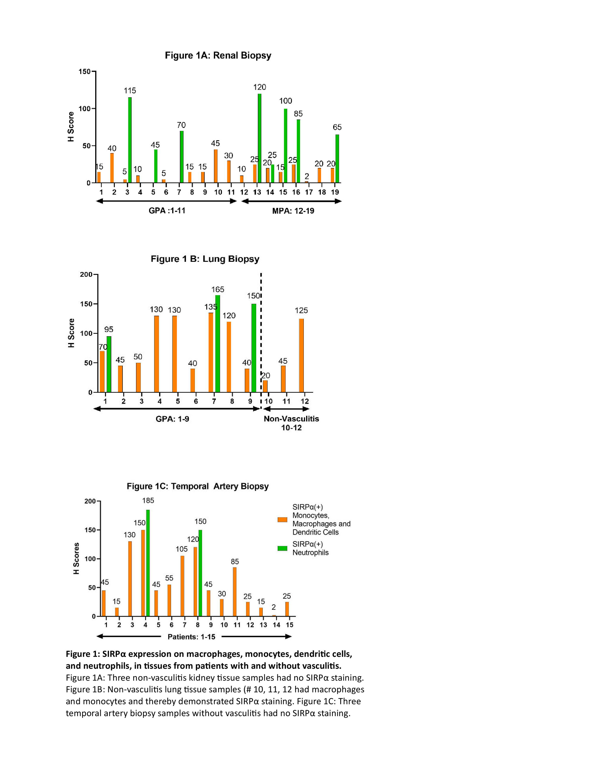
Figure 1A: Three non-vasculitis kidney tissue samples had no SIRPα staining. Figure 1B: Non-vasculitis lung tissue samples (# 10, 11, 12 had macrophages and monocytes and thereby demonstrated SIRPα staining. Figure 1C: Three temporal artery biopsy samples without vasculitis had no SIRPα staining.

Supporting image 2

Figure 2: Tissue samples from patients with granulomatosis with polyangiitis, microscopic polyangiitis, and giant cell arteritis demonstrating SIRPα Expression. Figure: 2A-C: Renal biopsy from a patient with granulomatosis with polyangiitis; Figure 2D-F: Lung biopsy from a patient with granulomatosis with polyangiitis: Figure 2G-I: Temporal artery biopsy from a patient with giant cell arteritis. Figures A, D, and G: H&E staining; Figures B, E, and H: Control rabbit IgG monoclonal antibody (DA1E) stain which is the negative control stain for anti- SIRPα stain; Figures C, F , and I: anti-SIRPα antibody D613M stain. The yellow arrows demonstrate some areas of positive SIRPα stain. Magnification of all images was at 20X.

Supporting image 3

Figure 3: Density of infiltration of macrophages, monocytes, dendritic cells,
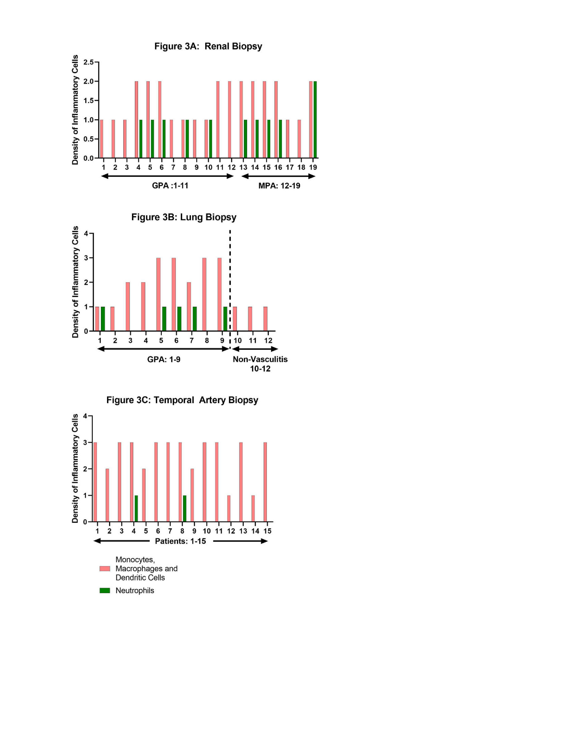
and neutrophils, in tissues from patients with and without vasculitis. An ordinal scale ranging from 0 to 3+ (0 = none, 1+ = rare/mild, 2+ = moderate, 3+ = severe/dense inflammatory infiltrate) was used to represent the density of each cell type infiltrate. Figure 3A: Three non-vasculitis kidney tissue samples had no inflammatory infiltrate. Figure 1B: Non-vasculitis lung tissue samples (# 10, 11, 12) had infiltration of macrophages and monocytes. Figure 1C: Three temporal artery biopsy samples without vasculitis had no inflammatory infiltrate.


Disclosures: S. Banerjee: None; E. Rose: Electra Therapeutics, Inc., 11, Star Therapeutics, Inc., 3, 11; S. Panicker: Electra Therapeutics, Inc., 11, Star Therapeutics, Inc., 3, 11; N. Khalidi: AbbVie/Abbott, 5, Bristol-Myers Squibb(BMS), 5, GlaxoSmithKlein(GSK), 1, Otsuka, 1, 6, Roche, 1, 6; C. Koening: Amgen, 1; C. Langford: AbbVie, 12, Non-paid consultant, AstraZeneca, 5, 12, Non-paid consultant, Bristol-Myers Squibb(BMS), 5, 12, Non-paid consultant, GlaxoSmithKlein(GSK), 5; P. Monach: Genentech, 12, Lecture with honorarium, HI-Bio, 2; C. Pagnoux: AstraZeneca, 1, 2, 6, GlaxoSmithKlein(GSK), 1, 6, Otsuka, 1, 2, 5, 6, Pfizer, 5, Roche, 2; C. McAlear: None; P. Merkel: AbbVie/Abbott, 5, Amgen, 2, 5, ArGenx, 2, AstraZeneca, 2, 5, Boehringer-Ingelheim, 2, 5, Bristol-Myers Squibb(BMS), 2, 5, Cabaletta, 2, CSL Behring, 2, Eicos, 5, Electra, 5, Genentech, 5, GlaxoSmithKlein(GSK), 2, 5, HiBio, 2, InflaRx, 2, 5, Janssen, 2, Jubilant, 2, Kyverna, 2, 11, MiroBio, 2, Neutrolis, 5, Novartis, 2, NS Pharma, 2, Q32, 2, Regeneron, 2, Sanofi, 2, Sparrow, 2, Takeda, 2, 5, UpToDate, 9, Visterra, 2.

To cite this abstract in AMA style:

Banerjee S, Rose E, Panicker S, Khalidi N, Koening C, Langford C, Monach P, Pagnoux C, McAlear C, Merkel P. SIRPα Expression in Systemic Vasculitis [abstract]. Arthritis Rheumatol. 2023; 75 (suppl 9). https://acrabstracts.org/abstract/sirp%ce%b1-expression-in-systemic-vasculitis/. Accessed .
  • Tweet
  • Click to email a link to a friend (Opens in new window) Email
  • Click to print (Opens in new window) Print

« Back to ACR Convergence 2023

ACR Meeting Abstracts - https://acrabstracts.org/abstract/sirp%ce%b1-expression-in-systemic-vasculitis/

Advanced Search

Your Favorites

You can save and print a list of your favorite abstracts during your browser session by clicking the “Favorite” button at the bottom of any abstract. View your favorites »

All abstracts accepted to ACR Convergence are under media embargo once the ACR has notified presenters of their abstract’s acceptance. They may be presented at other meetings or published as manuscripts after this time but should not be discussed in non-scholarly venues or outlets. The following embargo policies are strictly enforced by the ACR.

Accepted abstracts are made available to the public online in advance of the meeting and are published in a special online supplement of our scientific journal, Arthritis & Rheumatology. Information contained in those abstracts may not be released until the abstracts appear online. In an exception to the media embargo, academic institutions, private organizations, and companies with products whose value may be influenced by information contained in an abstract may issue a press release to coincide with the availability of an ACR abstract on the ACR website. However, the ACR continues to require that information that goes beyond that contained in the abstract (e.g., discussion of the abstract done as part of editorial news coverage) is under media embargo until 10:00 AM ET on November 14, 2024. Journalists with access to embargoed information cannot release articles or editorial news coverage before this time. Editorial news coverage is considered original articles/videos developed by employed journalists to report facts, commentary, and subject matter expert quotes in a narrative form using a variety of sources (e.g., research, announcements, press releases, events, etc.).

Violation of this policy may result in the abstract being withdrawn from the meeting and other measures deemed appropriate. Authors are responsible for notifying colleagues, institutions, communications firms, and all other stakeholders related to the development or promotion of the abstract about this policy. If you have questions about the ACR abstract embargo policy, please contact ACR abstracts staff at [email protected].

Wiley

  • Online Journal
  • Privacy Policy
  • Permissions Policies
  • Cookie Preferences

© Copyright 2025 American College of Rheumatology