Session Information
Title: Systemic Sclerosis, Fibrosing Syndromes, and Raynaud’s - Clinical Aspects and Therapeutics II
Session Type: Abstract Submissions (ACR)
Background/Purpose: Systemic sclerosis (SSc) is a multi-system disease with both visceral and cutaneous fibrosis. Dermal elasticity is reduced and stiffness increased due to excessive dermal and subcutaneous deposition of collagen leading to increased skin thickness as well as hardening and tightening of the skin. Shear wave elastography (SWE) is an emerging, operator-independent technique, which can obtain quantitative stiffness values. We describe a novel quantitative approach for evaluating the skin in scleroderma using shear wave elastography
Methods: We evaluated the skin of thirteen patients with diffuse systemic sclerosis and ten healthy controls using an Aixplorer shear wave ultrasound instrument (SupersonicImagine, Bothell, WA). Each patient underwent a SWE exam of the right arm at multiple sites (3rd finger, extensor forearm, upper arm), sternum, and face. The images consist of a spectrum of translucent colored bands superimposed on B-mode ultrasonographic images indicative of soft and highly elastic tissue to hard and minimally elastic tissue. The mean stiffness values of skin (measured in kPa units) were obtained at each site by averaging 3 measurements made per image obtained.
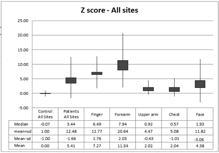
Results: The skin of seven men and five women with diffuse systemic sclerosis as well as 10 healthy controls was evaluated. The mean and 1SD SWE stiffness measures were (in kPa): finger –SSc 98.3 (SD-56) vs control – 32.1, (SD- 11.8); forearm – SSc 89.6 (SD- 58.9) vs control – 18.3 (SD-6.9), upper arm – SSc 34.5 (SD-19.2) vs control 17.7(SD8.6), face: SSc 19.3 (SD-17.7) vs control 8.4(SD-2.7), chest –SSc 23.6 (SD-17.1) vs control 10.9(SD-6.0). Maximum (out of range high) values of 300kPA were obtained for 1 patient at the finger and two patients at the forearm. Two-tailed t –tests were performed between the means of patients and controls at each site. Statistical significance (p<0.01) was achieved at the finger, forearm, all sites combined, and (p<0.05) at the upper arm. Statistical significance was not reached for the chest (p=0.056) and the face (p=0.08). Z scores (based on the mean and SD of each site in controls) were calculated for patient measurements at each individual site and combined sites compared to controls. Z scores at all sites were above the mean for controls (see figure 1).
Conclusion: Shear wave elastography is a valid and feasible quantitative method for assessing abnormal elasticity of the skin in scleroderma patients. It adds a new dimension to the assessment of skin involvement in SSc, and is an objective, non-invasive tool for potential use in clinical trials. Studies comparing its performance to stage of disease, skin thickness, and mRss are ongoing.
Disclosure:
I. Sacksen,
None;
M. H. Wener,
None;
P. S. Pollock,
None;
M. K. Dighe,
None.
« Back to 2013 ACR/ARHP Annual Meeting
ACR Meeting Abstracts - https://acrabstracts.org/abstract/shear-wave-elastography-a-novel-quantitative-approach-for-evaluating-scleroderma-skin/

