ACR Meeting Abstracts

ACR Meeting Abstracts

  • Meetings
    • ACR Convergence 2024
    • ACR Convergence 2023
    • 2023 ACR/ARP PRSYM
    • ACR Convergence 2022
    • ACR Convergence 2021
    • ACR Convergence 2020
    • 2020 ACR/ARP PRSYM
    • 2019 ACR/ARP Annual Meeting
    • 2018-2009 Meetings
    • Download Abstracts
  • Keyword Index
  • Advanced Search
  • Your Favorites
    • Favorites
    • Login
    • View and print all favorites
    • Clear all your favorites
  • ACR Meetings

Abstract Number: 485

Sexual Dimorphism of Popliteal Lymph Node Collapse As a Biomarker of Disease Progression in the Tumor Necrosis Factor Transgenic Mouse Model of Rheumatoid Arthritis

Emily Wu1, Richard Bell2, Christopher Rudmann3, Ronald Wood4, Christopher T. Ritchlin5, Homaira Rahimi6 and Edward Schwarz7, 1Department of Immunology, Microbiology, and Virology, University of Rochester, Rochester, NY, 2Pathology, University of Rochester, Rochester, NY, 3Biological Sciences, Carnegie Mellon University, Pittsburgh, PA, 4University of Rochester, Rochester, NY, 5Allergy Immunology & Rheumatology, University of Rochester Medical Center, Rochester, NY, 6Rheumatology, University of Rochester/Golisano Children's Hosp, Rochester, NY, 7Orthopedeatrics, University of Rochester, Rochester, NY

Meeting: 2016 ACR/ARHP Annual Meeting

Date of first publication: September 28, 2016

Keywords: animal models and ultrasound, rheumatoid arthritis

  • Tweet
  • Click to email a link to a friend (Opens in new window) Email
  • Click to print (Opens in new window) Print
Session Information

Date: Sunday, November 13, 2016

Title: Rheumatoid Arthritis – Animal Models - Poster I

Session Type: ACR Poster Session A

Session Time: 9:00AM-11:00AM

Background/Purpose: Although rheumatoid arthritis (RA) tends to occur earlier and with increased severity in females, the underling etiology of this sexual dimorphism is unknown. We have investigated the behavior of draining lymph nodes in murine models of inflammatory-erosive arthritis as a potential translational biomarker of arthritic initiation and flare. We have shown in tumor necrosis factor-transgenic (TNF-Tg) mice that ankle arthritis commences with enlargement of the popliteal lymph node (PLN) that continues to expand until the volume suddenly and stochastically collapses, coinciding with arthritic flare in the ipsilateral knee. To assess if this biomarker of disease onset and progression recapitulates the sexual dimorphism observed in clinical RA, we performed a longitudinal power Doppler ultrasound (PD-US) study of PLN in male and female TNF-Tg mice.  

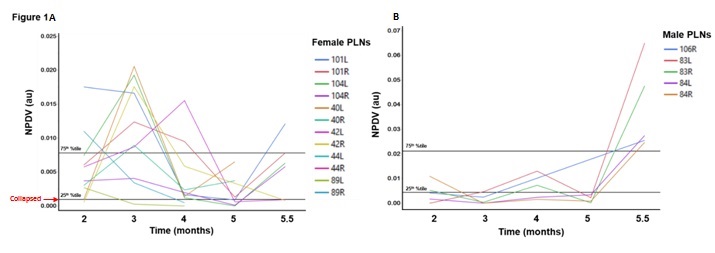
Methods:   PD-US was performed on the PLN and knee of each leg in male (n=3 mice; 5 PLN/knees) and female (n=6 mice; 12 PLN/knees) TNF-Tg mice at 2, 3, 4, 5, and 5.5 months of age. PLNs were phenotyped as expanding if the normalized PD (NPDV) signal exceeded the 75th percentile, or collapsed if it dropped below the 25th percentile after an expanding classification, as previously described (1). Each PLN/knee was evaluated independently, and percentiles were computed for each sex separately. The frequencies of expanded or collapsed PLN over time were compared using Fisher’s exact test; and knee NPDV comparisons used RM-ANOVA.  

Results: There were more expanded nodes at 3 months in female TNF-Tg mice (Figure 1a) compared to males (Figure 1b) (6 vs 0, p<0.01), but at 5.5-months, male animals had significantly more expanding nodes (5 vs 0, p<0.01).  The transition to collapsed nodes occurred in 5 females during the study period compared to 0 collapsed in males. Even more strikingly, all of the females with collapsed nodes before 4 months of age died before the final 5.5 month time point. There were no significant differences between sexes in knee NPDV at any time point, however both female and male displayed significantly increased NPDV at 5.5 vs. 5 months of age (0.19 ± 0.10, 0.24±0.04  vs 0.04 ± 0.04, 0.11 ± 0.10, p<0.05).

Conclusion: This is the first demonstration that PLN collapse occurs earlier in female TNF-Tg mice. Interestingly, knee NPDV increases, indicative of inflammatory-erosive arthritis, occur at the same time in both sexes. Taken together, this data supports the hypothesis that the mechanisms of RA are sex dependent, confirming this animal model as representative of the sexual dimorphism seen in patients. Further work is needed to elucidate the mechanism for early PLN collapse and mortality of female TNF-Tg mice. 1.         Bouta EM, Ju Y, Rahimi H, de Mesy-Bentley KL, Wood RW, Xing L, et al. Power Doppler Ultrasound Phenotyping of Expanding versus Collapsed Popliteal Lymph Nodes in Murine Inflammatory Arthritis. PLoS One. 2013;8(9):e73766.


Disclosure: E. Wu, None; R. Bell, None; C. Rudmann, None; R. Wood, None; C. T. Ritchlin, Amgen, Janssen Pharmaceutica Product, L.P., and UCB, 2,AbbVie, Amgen, Janssen Pharmaceutica Product, L.P., Regeneron, and UCB, 5; H. Rahimi, None; E. Schwarz, None.

To cite this abstract in AMA style:

Wu E, Bell R, Rudmann C, Wood R, Ritchlin CT, Rahimi H, Schwarz E. Sexual Dimorphism of Popliteal Lymph Node Collapse As a Biomarker of Disease Progression in the Tumor Necrosis Factor Transgenic Mouse Model of Rheumatoid Arthritis [abstract]. Arthritis Rheumatol. 2016; 68 (suppl 10). https://acrabstracts.org/abstract/sexual-dimorphism-of-popliteal-lymph-node-collapse-as-a-biomarker-of-disease-progression-in-the-tumor-necrosis-factor-transgenic-mouse-model-of-rheumatoid-arthritis/. Accessed .
  • Tweet
  • Click to email a link to a friend (Opens in new window) Email
  • Click to print (Opens in new window) Print

« Back to 2016 ACR/ARHP Annual Meeting

ACR Meeting Abstracts - https://acrabstracts.org/abstract/sexual-dimorphism-of-popliteal-lymph-node-collapse-as-a-biomarker-of-disease-progression-in-the-tumor-necrosis-factor-transgenic-mouse-model-of-rheumatoid-arthritis/

Advanced Search

Your Favorites

You can save and print a list of your favorite abstracts during your browser session by clicking the “Favorite” button at the bottom of any abstract. View your favorites »

All abstracts accepted to ACR Convergence are under media embargo once the ACR has notified presenters of their abstract’s acceptance. They may be presented at other meetings or published as manuscripts after this time but should not be discussed in non-scholarly venues or outlets. The following embargo policies are strictly enforced by the ACR.

Accepted abstracts are made available to the public online in advance of the meeting and are published in a special online supplement of our scientific journal, Arthritis & Rheumatology. Information contained in those abstracts may not be released until the abstracts appear online. In an exception to the media embargo, academic institutions, private organizations, and companies with products whose value may be influenced by information contained in an abstract may issue a press release to coincide with the availability of an ACR abstract on the ACR website. However, the ACR continues to require that information that goes beyond that contained in the abstract (e.g., discussion of the abstract done as part of editorial news coverage) is under media embargo until 10:00 AM ET on November 14, 2024. Journalists with access to embargoed information cannot release articles or editorial news coverage before this time. Editorial news coverage is considered original articles/videos developed by employed journalists to report facts, commentary, and subject matter expert quotes in a narrative form using a variety of sources (e.g., research, announcements, press releases, events, etc.).

Violation of this policy may result in the abstract being withdrawn from the meeting and other measures deemed appropriate. Authors are responsible for notifying colleagues, institutions, communications firms, and all other stakeholders related to the development or promotion of the abstract about this policy. If you have questions about the ACR abstract embargo policy, please contact ACR abstracts staff at [email protected].

Wiley

  • Online Journal
  • Privacy Policy
  • Permissions Policies
  • Cookie Preferences

© Copyright 2025 American College of Rheumatology