Session Information
Session Type: ACR Poster Session A
Session Time: 9:00AM-11:00AM
Background/Purpose:
PET imaging with macrophage tracers has been shown promising for detection of (sub)clinical synovitis, making it useful for both early diagnostics and therapy monitoring in rheumatoid arthritis (RA) patients (1,2). For detection of more subtle arthritis, a previously investigated macrophage tracer, (R)-[11C]PK11195, was limited in use due to high background uptake in bone and bone marrow. [18F]fluoro-PEG-folate binds to the folate receptor β, which is expressed on synovial macrophages (3). Preclinical research in arthritic rats has shown excellent targeting, making it a promising tracer for clinical testing in RA patients (4). In this study, we investigated the value of [18F]fluoro-PEG-folate PET-CT for imaging of inflamed joints in patients with clinically active RA.
Methods:
Nine RA patients with a minimal of two clinically inflamed hand joints were included. PET-CT scans were performed of the hands, after intravenous administration of either 185 MBq of [18F]fluoro-PEG-folate (n = 6) or 425 MBq of (R)-[11C]PK11195 (n = 3) for comparison. Joints with visually marked uptake were further analyzed by calculation of Standardized Uptake Values (SUVs) in Volumes of Interest (VOI), drawn on top of the PET positive joints. We drew background VOIs on metacarpal bone in order to calculate Target-to-Background (T/B) ratios.
Results:
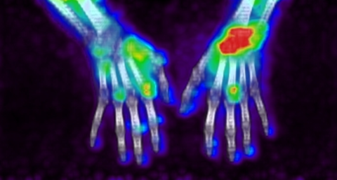
None of the patients showed adverse effects, establishing the safety of [18F]fluoro-PEG-folate for use in humans. Arthritic joints were clearly visualized (Fig 1).. In the patients injected with [18F]fluoro-PEG-folate, 25 positive joints were observed, with a minimum of two joints per patient. In 10 of these 25 joints, clinical arthritis was confirmed. In the remaining 15 positive joints clinical inflammation was absent, suggesting the presence of subclinical inflammation. Whilst both [18F]fluoro-PEG-folate and (R)-[11C]PK11195 showed clear uptake in arthritic joints, [18F]fluoro-PEG-folate demonstrated a significantly lower background uptake than (R)-[11C]PK11195 (SUV of 0.18 vs 0.75; p < 0.001) respectively. T/B-ratios were significantly higher for [18F]fluoro-PEG-folate (3.60vs 1.72, p = 0.009).
Figure 1: [18F]fluoro-PEG-folate uptake on PET-CT in inflamed hand/wrist joints of a RA patient.
Conclusion:
[18F]fluoro-PEG-folate shows great potential as a macrophage tracer to image both clinical and presumably also subclinical arthritis in RA patients. The tracer shows improved characteristics compared to the established macrophage tracer (R)-[11C]PK11195 for imaging arthritis, because of lower background signal.
References:
(1) Gent YY, et al. J Rheumatology. 2014; 41: 2145-52
(2) Gent YY, et al. Arthritis Rheum. 2012; 64: 62-6
(3) Chandrupatla DMSH, Arthritis Res Ther. 2017; 19: 114.
(4) Gent YY, et al. Arthritis Res Ther. 2013; 15: R37
To cite this abstract in AMA style:
Verweij N, Bruijnen S, Gent Y, Huisman M, Jansen G, Molthoff C, Chen Q, Low P, Windhorst A, Lammertsma A, Hoekstra O, Voskuyl A, van der Laken C. Rheumatoid Arthritis Imaging on PET-CT Using a Novel Folate Receptor Ligand for Macrophage Targeting [abstract]. Arthritis Rheumatol. 2017; 69 (suppl 10). https://acrabstracts.org/abstract/rheumatoid-arthritis-imaging-on-pet-ct-using-a-novel-folate-receptor-ligand-for-macrophage-targeting/. Accessed .« Back to 2017 ACR/ARHP Annual Meeting
ACR Meeting Abstracts - https://acrabstracts.org/abstract/rheumatoid-arthritis-imaging-on-pet-ct-using-a-novel-folate-receptor-ligand-for-macrophage-targeting/

