Session Information
Session Type: Poster Session (Sunday)
Session Time: 9:00AM-11:00AM
Background/Purpose: Chronic systemic inflammation in patients with ankylosing spondylitis (AS) contributes to the development of cardiovascular disease (CVD). Early recognition of microvascular changes could improve prevention. The retinal vasculature has been proven to reflect systemic (micro)vascular changes. Narrowing of retinal arterioles, widening of retinal venules and decreased vascular density are associated with coronary artery disease. Furthermore, systemic inflammation was reported to result in wider retinal venules. Remarkably, studies in AS patients are lacking, while the retinal vasculature is easily accessible and could provide an opportunity to recognize preclinical CVD. This study compared the retinal microvasculature of AS patients to healthy controls, and its association with cardiovascular risk.
Methods: Cross-sectional case-control study comparing AS patients with healthy controls from a cohort of the Dutch Twins Register.(1) Inclusion criteria were: age 50-75 years and no history of diabetes mellitus or cerebrovascular disease. AS patients fulfilled the modified New York criteria. Controls with an auto-inflammatory disease were excluded. Consecutive AS patients were recruited from the Rheumatology outpatient clinic of Reade and Amsterdam UMC (VUmc). Controls were selected from the pre-existing EMIF-AD PreClinAD cohort.(1) All subjects underwent Optical Coherence Tomography Angiography (OCTA) and fundus photography, analyzed with Singapore I Vessel Assessment software (Table 1). Data on patient- and disease characteristics, CVD risk factors, medication and lipids were collected. For subjects without a previous CVD, the 10 years cardiovascular comorbidity/mortality risk was calculated (Systematic Coronary Risk Evaluation, SCORE). Data were evaluated with linear regression, correcting for age when comparing patients and controls. The association between the SCORE and ocular parameters was assessed stratified for patients and controls.
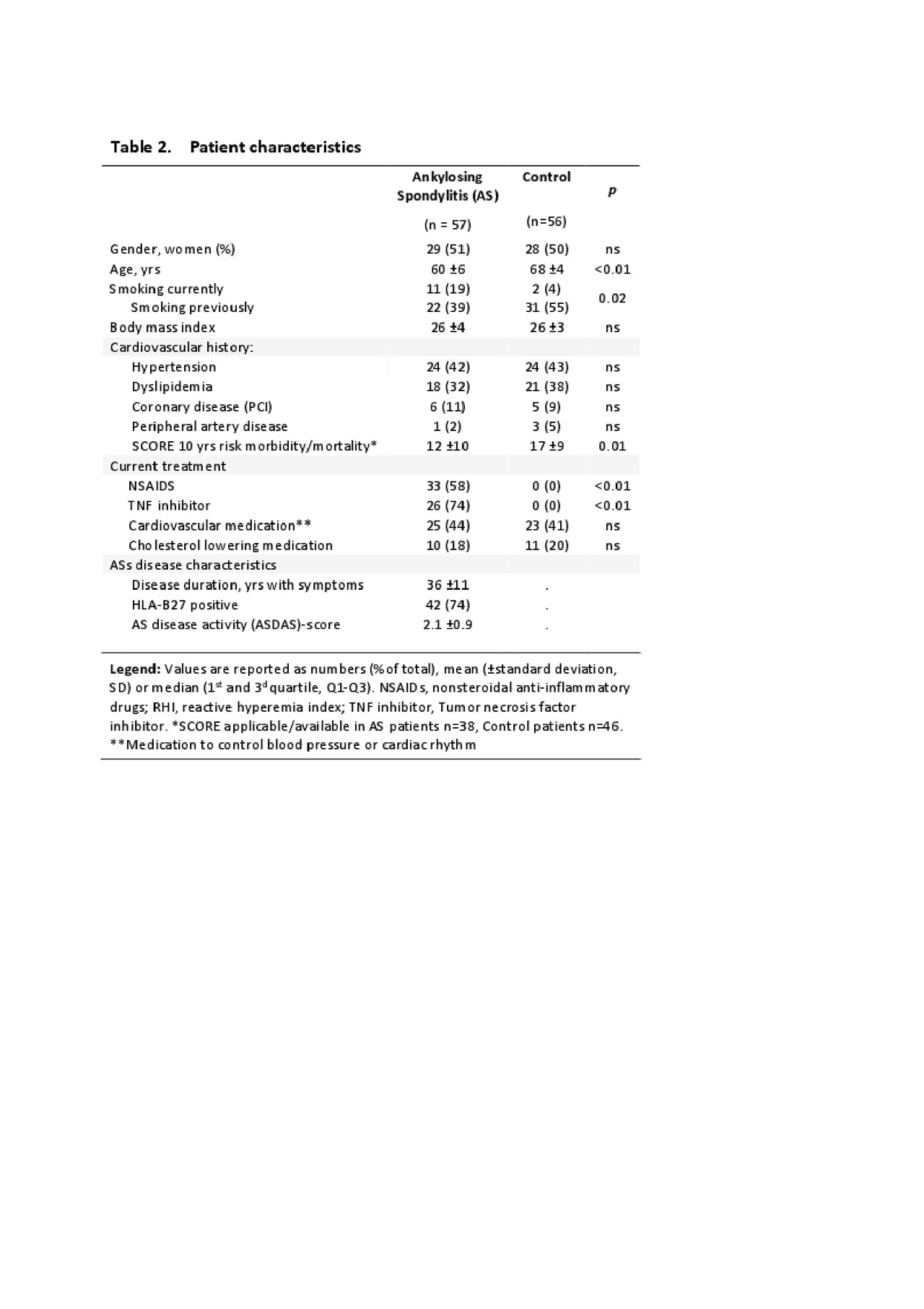
Results: In total, 57 AS patients (51% women) and 56 controls (50% women) were included. Both groups differed significantly in mean age (AS: 60±SD6 years; Controls 68±SD4; p< 0.01, Table 2). Fundus photo parameters (Table 1), in particular retinal arteriole and venule diameter, did not differ between patients and controls. However, patients showed significantly lower retinal vessel density (OCTA) compared to controls (age-corrected Beta: -0.53, p=0.046). Only in AS patients, lower retinal vessel density (p=0.02), arteriole fractal dimension; (p=.04) and arteriole diameter (p=.02) were associated with a higher cardiovascular risk (SCORE), but not in controls.
Conclusion: This study did not find differences in arteriolar and venular diameter between AS patients and controls. However, it is the first to report a significantly decreased retinal vessel density in AS patients, which was associated with a higher cardiovascular risk. Therefore, decreased vascular density might be an early sign of preclinical cardiovascular disease in AS patients.
Reference
- Konijnenberg E et al. The EMIF-AD PreclinAD study: study design and baseline cohort overview. Alzheimers Res Ther. 2018; 10:75.

Table 1 – Overview ocular vascular parameters

Table 2 – Patient characteristics
To cite this abstract in AMA style:
van Bentum R, Baniaamam M, Kinaci-Tas B, van de Kreeke A, Kocyigit M, Visser P, Serné E, Verbraak F, Nurmohamed M, van der Horst-Bruinsma I. Retina of Ankylosing Spondylitis Patients Shows Early Signs of Atherosclerotic Disease in Comparison with Healthy Controls [abstract]. Arthritis Rheumatol. 2019; 71 (suppl 10). https://acrabstracts.org/abstract/retina-of-ankylosing-spondylitis-patients-shows-early-signs-of-atherosclerotic-disease-in-comparison-with-healthy-controls/. Accessed .« Back to 2019 ACR/ARP Annual Meeting
ACR Meeting Abstracts - https://acrabstracts.org/abstract/retina-of-ankylosing-spondylitis-patients-shows-early-signs-of-atherosclerotic-disease-in-comparison-with-healthy-controls/
