Session Information
Date: Monday, November 14, 2016
Title: Spondylarthropathies Psoriatic Arthritis – Pathogenesis, Etiology - Poster I
Session Type: ACR Poster Session B
Session Time: 9:00AM-11:00AM
Background/Purpose: Earlier we have reported that synovial tissues of psoriatic arthritis (PsA) and rheumatoid arthritis (RA) are enriched with the Th9 cells along with its key cytokine IL-9 (1). Here we are reporting new functions of IL-9; its regulatory role on (i) FLS (fibroblast like synoviocyte) biology and (ii) pannus formation in PsA and RA.
Methods : FLS were isolated from synovial tissue biopsies of PsA, RA and OA (n=5, for each) and cultured (1). IL-9R expression was determined by immunobloting and RT-PCR studies. MTT assay and apoptosis assay (Annexin-V) were performed to determine the pro-growth/survival effect of rIL-9 on FLS. In the cultured FLS IL-6, IL-8, MMP-3 were measured by ELISA.
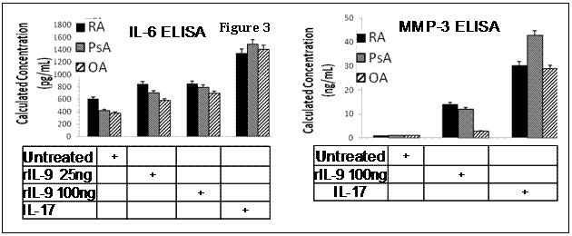
Results: RT-PCR and immunoblot studies demonstrated presence of IL9-R in FLS of PsA, RA and OA (Fig 1). rIL-9 induced proliferation of FLS and inhibited TNF-a induced apoptosis (Fig 2). rIL-9 induced IL-6 and MMP-3 expression in FLS (Fig 3).
Conclusion: Here we observed that FLS survival/prolifeartion, inflammatory cytokine expression and upregulation of MMP 3 the major components of the inflammatory proliferative cascades for pannus formation are regulated by IL-9. This is the first report to identify the presence of IL-9R on FLS and the functional significance of the IL-9/IL-9R system on FLS biology in PsA and RA. Figure 1. IL-9R is expressed in FLS – IL-9R gene was expressed in FLS of PsA and RA patients. Compared to the unactivated T lymphocytes the relative gene expression of IL-9R in FLS of PsA and RA patients was 3.5±0.67, 2.4±0.5, respectively (p<.01, Fig 1). IL-9R expression was confirmed by immunoblotting. The relative intensity (R.I) of IL-9R in FLS of PsA, RA and OA patients was 0.3 ± 0.02, 0.37 ± 0.05, 0.28 ± 0.08 respectively.
Figure 2. Human rIL-9 promoted survival of FLS– To test the functional significance of the IL9R we investigated whether rIL-9 can promote FLS survival. FLS of PsA, RA and OA were treated with rIL-9 (25 ng/ml) for 48hrs then TNFα (20 ng/ml) for 24 hours to induce apoptosis (1). In the FLS of PsA, TNFα + rIL-9 induced 11.41±3.61% apoptosis compared to 35±5.53% induced apoptosis seen in TNFα alone (p<.01). Similar results were seen in RA and OA FLS.
Figure 3. Compared to the untreated FLS, rIL-9 (100 ng/ml) induced upregulation of IL-6 and MMP- 3 (p<.01).
Reference: 1. Raychaudhuri SK, et al. Cytokine 2016; 79:45-51.
To cite this abstract in AMA style:
Raychaudhuri SP, Raychaudhuri SK. Regulatory Role of the IL-9/IL-9R System on Pannus Formation in Psoriatic Arthritis [abstract]. Arthritis Rheumatol. 2016; 68 (suppl 10). https://acrabstracts.org/abstract/regulatory-role-of-the-il-9il-9r-system-on-pannus-formation-in-psoriatic-arthritis/. Accessed .« Back to 2016 ACR/ARHP Annual Meeting
ACR Meeting Abstracts - https://acrabstracts.org/abstract/regulatory-role-of-the-il-9il-9r-system-on-pannus-formation-in-psoriatic-arthritis/


