Session Information
Title: Imaging of Rheumatic Diseases I: Imaging in Gout, Pediatric, Soft and Connective Tissue Diseases
Session Type: Abstract Submissions (ACR)
Background/Purpose: Blau syndrome (Blau) is a rare auto-inflammatory disease, and it has now been shown to be caused by NOD2/CARD15 gene mutations. Clinical features of Blau consist of granulomatous inflammation in the joints, eyes, and skin. However, most of the patients were initially miss-diagnosed as JIA because the symmetrical polyarthritis occurred in early phase of the disease, and “boutonniere” deformities-like fingers appeared during the disease course. However, the articular involvement in Blau has been considered as non-erosive. Therefore, we evaluate the joint damage in Blau patients by measuring calpal length (CL), an objective tool for evaluating cartilage damage in polyariticular JIA (poJIA)1-2).
Methods: A total of 6 Blau patients were followed up their CL for mean 9.2 years. Their disease duration at the first CL measurement was 5.2 years. Since biologic agents were used in 5 of 6 of the Blau patients, changes in CL of 46 poJIA patients treated with biologics were also followed up. CL was analyzed by standard deviation (SD) calculated by Poznanski’s formula established from healthy children3).
In addition, ultrasound examination was carried out in 4 Blau patients at the last observation. A total of 3 joints and 4 tenosynovial sites in the wrist were scanned and the ultrasound images of gray-scale (GS) or Power Doppler (PD) findings were graded from 0 (normal) to 3 (severe).
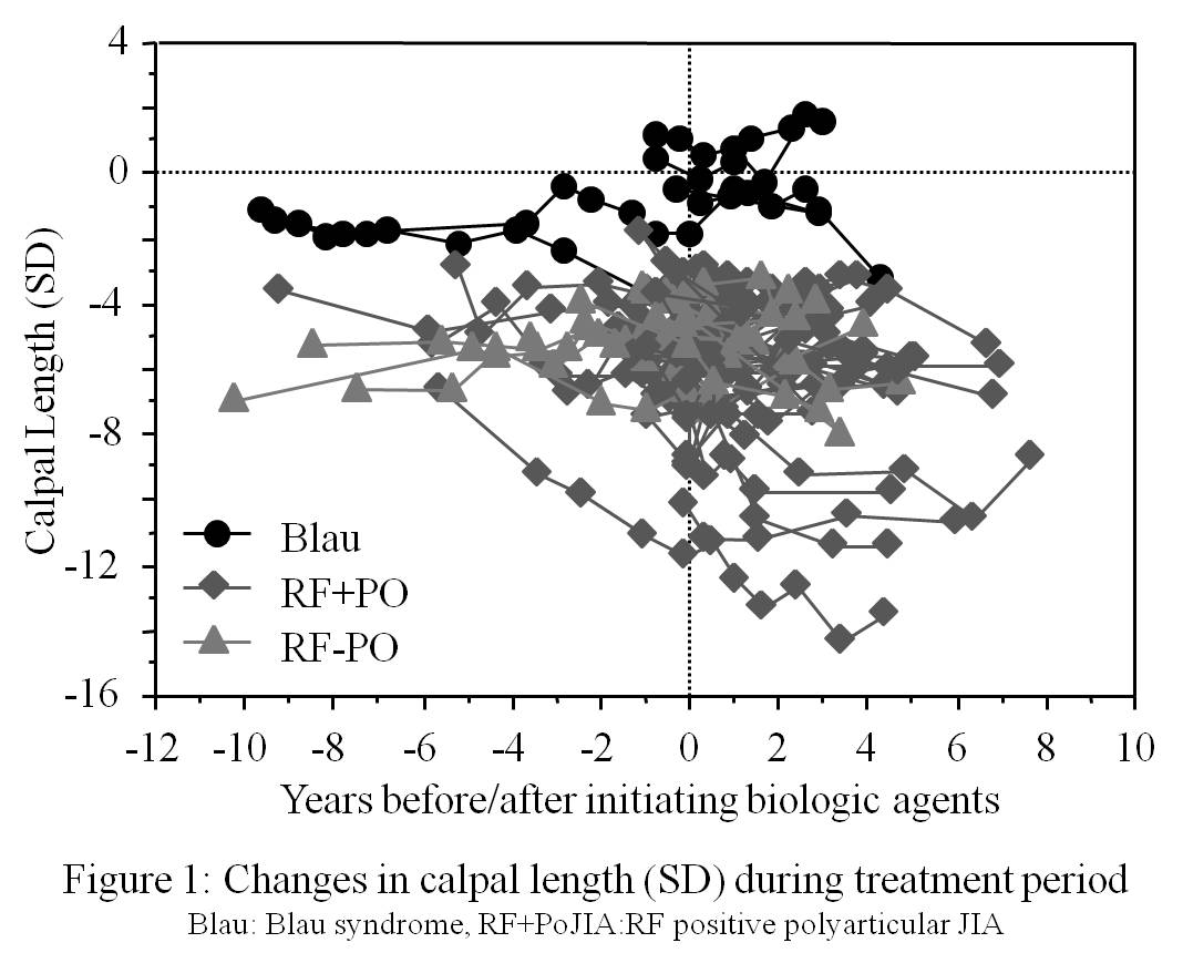 Results: Though all Blau patients had comptodactyly with “boutonniere” deformities like fingers, CL(SD) maintained normal range (within – 2SD to +2SD) during the observation period regardless of biologic therapy (Figure 1). On the contrary, CL(SD) in poJIA decreased with disease course especially before initiating biologic therapy, and there were negative correlation between the CL(SD) and the disease duration in both RF positive (p=0.0018) and RF negative (p=0.008) poJIA patients.
Ultrasound examination revealed that tenosynovitis was more frequent and severe than intra-articular synovitis in the 8 wrists of 4 Blau patients; Grade 1-3 findings were found in 62.5% by PD and 31.2% by GS in scanned tenosynovium sites, while it was 29.2% by PD and 4.2% by GS in intra-articular synovium sites (tendon vs intra-articular sites: p=0.0169 by PD, p=0.0161 by GS).
Conclusion: Comptodactyly with “boutonniere” deformities-like findings, a characteristic feature of Blau, may be caused by tenosynovitis but not by cartilage destruction in finger joints. The finding indicates the usefulness of CL and ultrasound examination in differentiating Blau from JIA, and is also suggestive that tenosynovitis may be more predominant than intra-articular synovitis in Blau syndrome.
1) Ravelli A, et al. J Pediatr 1998; 133:262-5. 2) Kubota T et al. #1163, 2012 ACR Annual Meeting. 3) Poznanski AK et al. Radiology 1978;129:661-8.
Disclosure:
T. Yamatou,
None;
T. Kubota,
None;
H. Akaike,
None;
Y. Yamasaki,
None;
Y. Nonaka,
None;
Y. Nerome,
None;
T. Takezaki,
None;
H. Imanaka,
None;
K. Ikeda,
None;
N. Kambe,
None;
S. Takei,
Chugai, Eisai, Takeda, and Bristol-Myers,
2,
Chugai, Eisai, Takeda, Abbvie, Astellas, Teijin, Novartis, Pfizer, and Asahi Kasei,
8;
T. Nagakura,
None.
« Back to 2013 ACR/ARHP Annual Meeting
ACR Meeting Abstracts - https://acrabstracts.org/abstract/quantitative-image-analysis-of-articular-involvement-in-blau-syndrome-by-radiographic-calpal-length-and-ultrasound-assessment/