ACR Meeting Abstracts

ACR Meeting Abstracts

  • Meetings
    • ACR Convergence 2024
    • ACR Convergence 2023
    • 2023 ACR/ARP PRSYM
    • ACR Convergence 2022
    • ACR Convergence 2021
    • ACR Convergence 2020
    • 2020 ACR/ARP PRSYM
    • 2019 ACR/ARP Annual Meeting
    • 2018-2009 Meetings
    • Download Abstracts
  • Keyword Index
  • Advanced Search
  • Your Favorites
    • Favorites
    • Login
    • View and print all favorites
    • Clear all your favorites
  • ACR Meetings

Abstract Number: 0895

Procoagulant Extracellular Vesicles in Patients with Livedo Reticularis

Avital Baniel1, Cristina Ricco2, Rohan Dhiman3, Ahmed Eldaboush4, Caroline Stone4, Daniella Faden4, Luca Musante4, MIng-lin Liu4 and Victoria Werth5, 1Perelman School of Medicine, UPENN, Philadelphia, PA, 2university of pennsylvania, Philadelphia, 3University of Pennsylvania, Newport Coast, CA, 4University of pennsylvania, Philadelphia, PA, 5University of Pennsylvania, Wynnewood, PA

Meeting: ACR Convergence 2024

Keywords: Dermatology, proteomics

  • Tweet
  • Email
  • Print
Session Information

Date: Sunday, November 17, 2024

Title: Genetics, Genomics & Proteomics Poster

Session Type: Poster Session B

Session Time: 10:30AM-12:30PM

Background/Purpose: Procoagulant activity is an important property of EVs. Connective tissue disease (CTD) patients with livedo reticularis (LR) are known for their procoagulant conditions. The objective of this study is to investigate the role of EVs in LR. 

Methods: We isolated EVs from plasma of 12 LR patients and 7 controls using sequential ultracentrifugation and size-exclusion chromatography. The EVs were analyzed by protein mass spectrometry. We measured tissue factor (TF) procoagulant activity and analyzed the cellular origin of EVs.

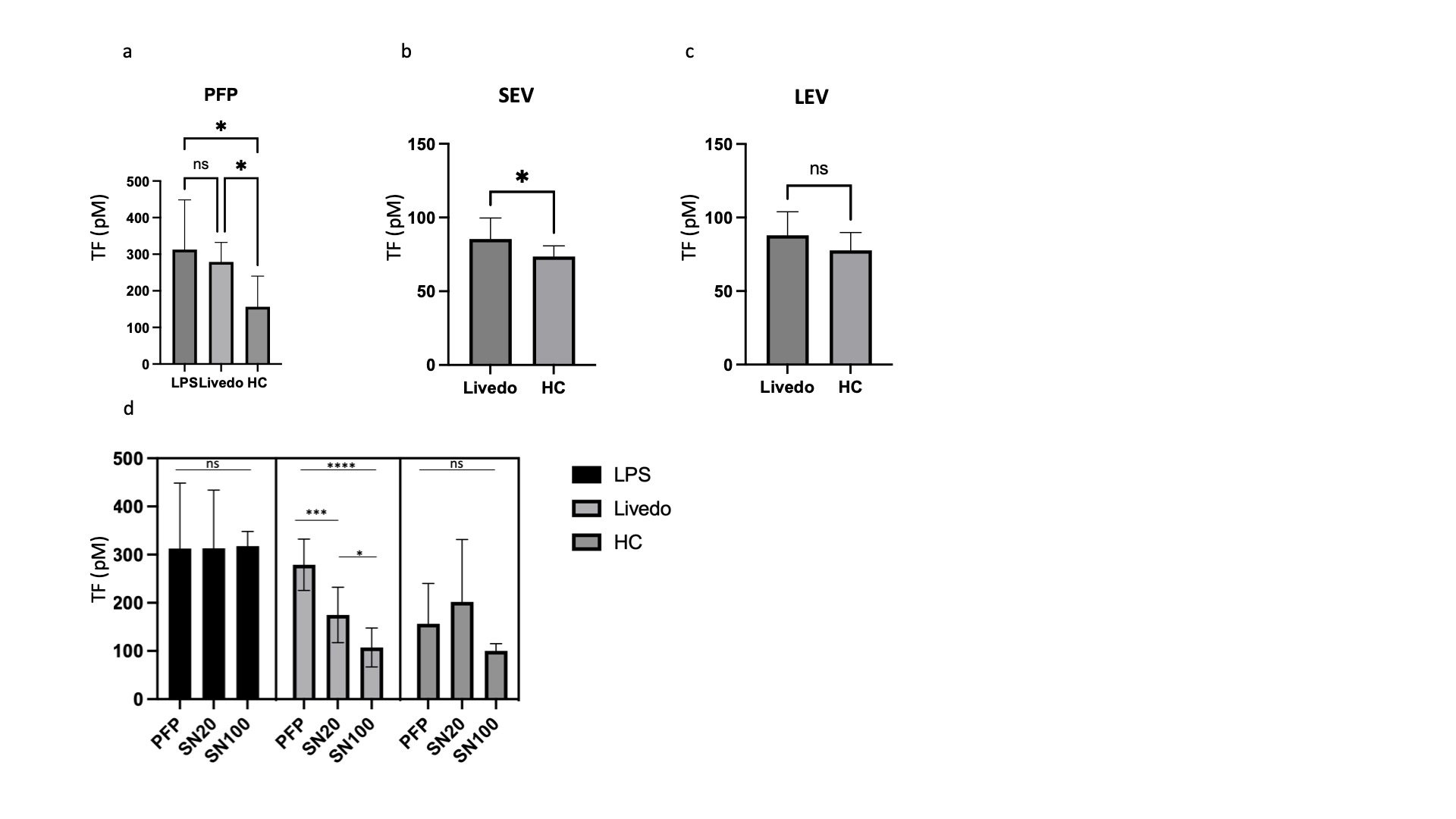
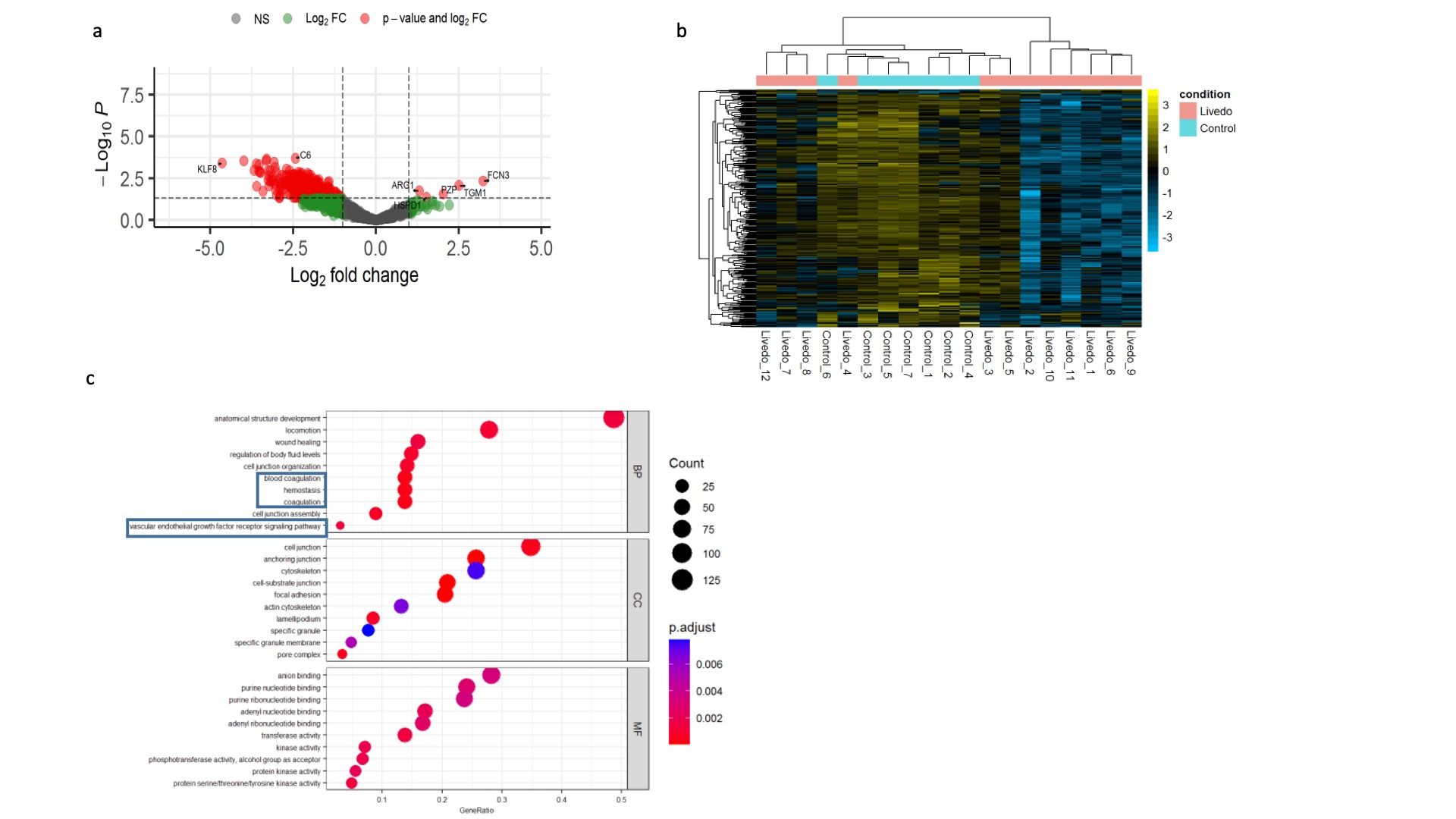
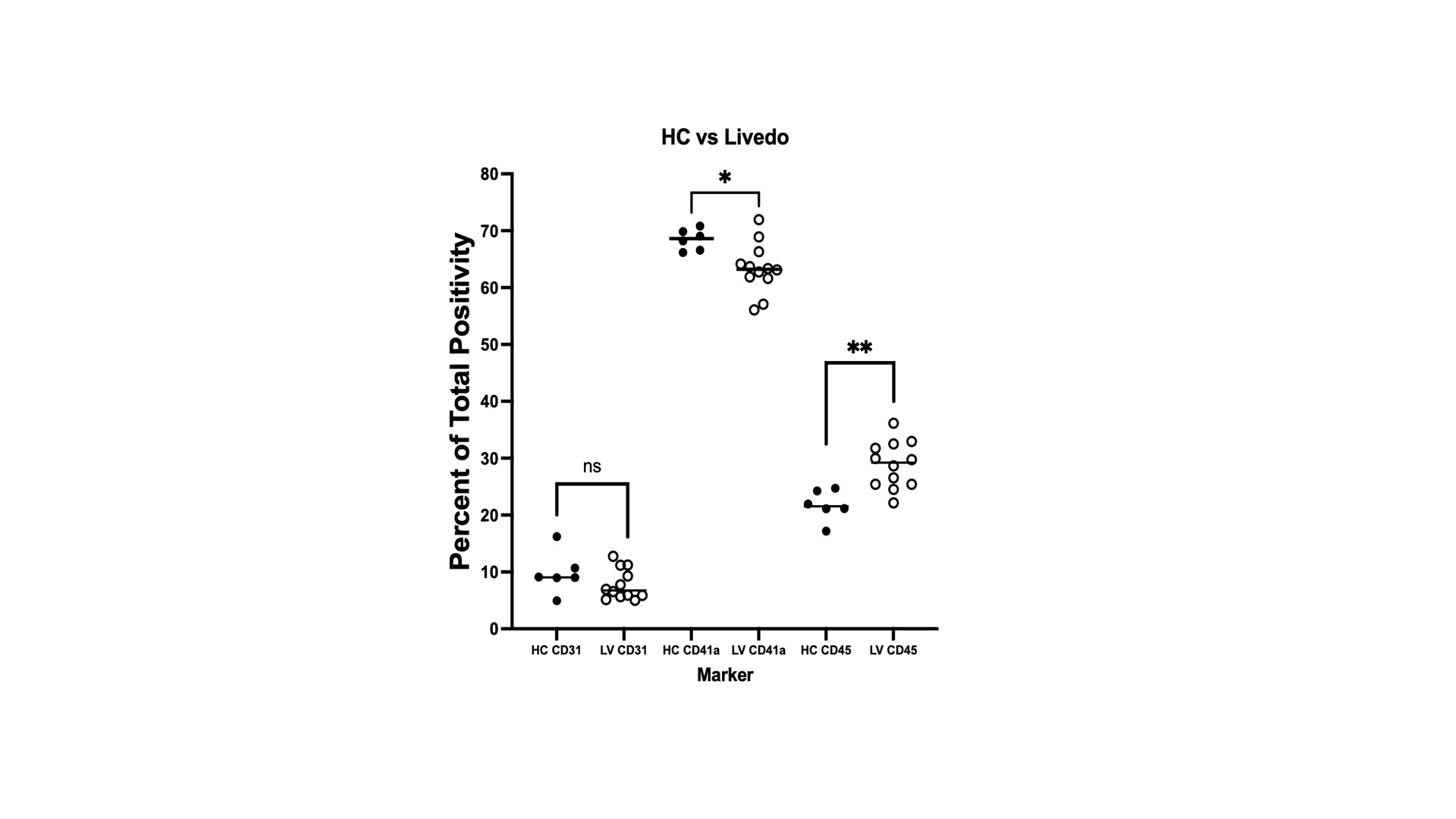
Results: TF procoagulant activity was increased in the plasma of LR patients compared to healthy controls (HCs). In EVs, we detected increased levels of TF activity in small EVs (SEV) but not in large EVs (LEV) in LR patients. In the process of sequential centrifugation, we measured TF activity in supernatants after each step. The 1st supernatant represented plasma without LEVs and the 2nd represented plasma depleted of both LEVs and SEVs. LR supernatants demonstrated a decrease in TF activity with each step, supporting a role for both large and small EVs in TF activation. This decrease was not seen in supernatants derived from HCs or from LPS-induced samples that were used as positive controls. We did not detect a difference in TF activity between large and small EVs. The concentration of vesicles in plasma was not increased in LR patients, implying the procoagulant properties of EVs may not be explained by a higher abundance, as reported in other procoagulant conditions, but possibly by their cargo. Hence, we sought to analyze the protein content of EVs. We detected 304 differentially expressed proteins (DEP) between LR and HCs. Five were upregulated and 269 downregulated. Most LR patients revealed a distinct expression pattern compared to HCs. A subset with profound vasculopathy showed a different pattern and for patients in remission the pattern was similar to HCs. Overrepresentation analysis revealed enrichment of proteins associated with homeostasis, coagulation, and vascular endothelial growth factor signaling pathways. Amongst the DEPs, Krueppel-Like Factor 8 (KLF8) exhibited significant prominence, demonstrating a substantial decrease in abundance by a fold change of 4.6 (p< 0.001). KLF8 is expressed in vascular endothelium and regulates angiogenesis and vessel contractility. Finally, we investigated the cells of origin of EVs in LR and controls previously identified as sources of procoagulant EVs. Our analysis revealed that patients with LR exhibited an increase in leukocyte-derived EVs and a decrease in platelet-derived EVs when compared with EVs derived from HCs. No significant changes were observed in the fraction of EVs that originated from endothelial cells. This may be indicative of a shift from baseline homeostasis in health to an inflammatory EV profile in LR.

Conclusion: EVs exert a procoagulant effect in LR. Increased TF activity and a shift in EV release from primarily platelet-driven to those originating from leukocytes are possible mechanisms underlying this effect. Our findings suggest that a reduction in KLF8 within EVs may be a key regulatory factor in LR.

Supporting image 1

Figure 1: Tissue factor (TF) activity. Plasma from Livedo reticularis (LR) and healthy controls (HC) was collected and sequentially centrifuged to prepare platelet free plasma (PFP) and extracellular vesicle (EV) preps. For positive control whole blood collected from HC and stimulated with LPS for 5 hours at 37 ◦C and processed in the same way to prepare PFP and EV preps. TF activity was measured by a chromogenic activity assay kit that quantifies the amount of FXa produced and is directly proportional to the TF enzymatic activity. TF activity in PFP was increased in both plasma of LR patients and positive controls compared to HCs (a). TF activity detected in SEVs is increased in LR patients compared to HCs (b) No significant change was detected in LEVs (c). Sequential filtration of particles decreased TF activity only in LR samples but not in HCs or positive controls (d). SN20 supernatant 20, represents plasma depleted of large particles. SN100 supernatant 100, represents plasma depleted of both large and small particles. Data represent means±SDs. *p<0.05, **p<0.01, ***p<0.001, ****p<0.0001 between groups as indicated.

Supporting image 2

Figure 2: Proteomic Analysis. EVs were subjected to proteomic analysis using mass spectrometry. Volcano plot demonstrating the distribution of differentially expressed proteins (a). The heatmap of protein cargo in EVs reveals a distinct profile for LR patients (b). Enrichment analysis based on the Gene Ontology database revealed notable associations with coagulation, homeostasis, and vascular endothelial growth factor receptor signaling pathways (c).

Supporting image 3

Figure 3: Cell of origin analysis. To investigate potential differences in the cellular origins of extracellular vesicles (EVs), flow cytometry was employed. Our analysis revealed that in LR patients, EV release was significantly elevated in leukocytes (indicated by CD45 expression) and reduced in platelets (indicated by CD41 expression) compared to healthy controls (HC). No significant variation was observed in the fluorescence intensity of the endothelial cell marker CD31.


Disclosures: A. Baniel: None; C. Ricco: None; R. Dhiman: None; A. Eldaboush: None; C. Stone: None; D. Faden: None; L. Musante: None; M. Liu: None; V. Werth: AbbVie/Abbott, 2, Alpine immune sciences, 2, Amgen, 1, 5, anaptysbio, 2, AstraZeneca, 2, 5, Biogen, 2, 5, BMS, 2, 5, Cabaletta Bio, 2, Calyx, 2, Caribou, 2, Corbus, 5, CSL Behring, 2, 5, Cugene, 2, Evommune, 2, Gilead, 2, 5, GSK, 2, Horizon, 2, 5, Immunovant, 2, Innovaderm, 2, Janssen, 2, Lilly, 2, Merck, 2, Nuvig Pharmaceuticals, 2, Pfizer, 2, 5, Priovant, 5, Regeneron, 1, 5, Rome Pharmaceuticals, 2, 5, Sanofi, 2, Takeda, 2, UCB, 2, Ventus, 2, 5, Viela, 5, Xencor, 2.

To cite this abstract in AMA style:

Baniel A, Ricco C, Dhiman R, Eldaboush A, Stone C, Faden D, Musante L, Liu M, Werth V. Procoagulant Extracellular Vesicles in Patients with Livedo Reticularis [abstract]. Arthritis Rheumatol. 2024; 76 (suppl 9). https://acrabstracts.org/abstract/procoagulant-extracellular-vesicles-in-patients-with-livedo-reticularis/. Accessed .
  • Tweet
  • Email
  • Print

« Back to ACR Convergence 2024

ACR Meeting Abstracts - https://acrabstracts.org/abstract/procoagulant-extracellular-vesicles-in-patients-with-livedo-reticularis/

Advanced Search

Your Favorites

You can save and print a list of your favorite abstracts during your browser session by clicking the “Favorite” button at the bottom of any abstract. View your favorites »

All abstracts accepted to ACR Convergence are under media embargo once the ACR has notified presenters of their abstract’s acceptance. They may be presented at other meetings or published as manuscripts after this time but should not be discussed in non-scholarly venues or outlets. The following embargo policies are strictly enforced by the ACR.

Accepted abstracts are made available to the public online in advance of the meeting and are published in a special online supplement of our scientific journal, Arthritis & Rheumatology. Information contained in those abstracts may not be released until the abstracts appear online. In an exception to the media embargo, academic institutions, private organizations, and companies with products whose value may be influenced by information contained in an abstract may issue a press release to coincide with the availability of an ACR abstract on the ACR website. However, the ACR continues to require that information that goes beyond that contained in the abstract (e.g., discussion of the abstract done as part of editorial news coverage) is under media embargo until 10:00 AM ET on November 14, 2024. Journalists with access to embargoed information cannot release articles or editorial news coverage before this time. Editorial news coverage is considered original articles/videos developed by employed journalists to report facts, commentary, and subject matter expert quotes in a narrative form using a variety of sources (e.g., research, announcements, press releases, events, etc.).

Violation of this policy may result in the abstract being withdrawn from the meeting and other measures deemed appropriate. Authors are responsible for notifying colleagues, institutions, communications firms, and all other stakeholders related to the development or promotion of the abstract about this policy. If you have questions about the ACR abstract embargo policy, please contact ACR abstracts staff at [email protected].

Wiley

  • Online Journal
  • Privacy Policy
  • Permissions Policies
  • Cookie Preferences

© Copyright 2025 American College of Rheumatology