ACR Meeting Abstracts

ACR Meeting Abstracts

  • Meetings
    • ACR Convergence 2024
    • ACR Convergence 2023
    • 2023 ACR/ARP PRSYM
    • ACR Convergence 2022
    • ACR Convergence 2021
    • ACR Convergence 2020
    • 2020 ACR/ARP PRSYM
    • 2019 ACR/ARP Annual Meeting
    • 2018-2009 Meetings
    • Download Abstracts
  • Keyword Index
  • Advanced Search
  • Your Favorites
    • Favorites
    • Login
    • View and print all favorites
    • Clear all your favorites
  • ACR Meetings

Abstract Number: 0062

Pro-Fibrotic Effects of Malondialdehyde-Acetaldehyde-Adducted and/or Citrullinated Proteins on Macrophages and Human Lung Fibroblasts

Lauren Klingemann1, Nozima Aripova1, Nigina Aripova2, Michael Duryee1, Carlos Hunter1, Jill Poole1, Bryant England1, Ted Mikuls1 and Geoffrey Thiele1, 1University of Nebraska Medical Center, Omaha, NE, 2WUSTL, St. Louis, MO

Meeting: ACR Convergence 2024

Keywords: citrullination, Collagen, Fibroblasts, Other, interstitial lung disease, macrophages

  • Tweet
  • Click to email a link to a friend (Opens in new window) Email
  • Click to print (Opens in new window) Print
Session Information

Date: Saturday, November 16, 2024

Title: RA – Etiology & Pathogenesis Poster

Session Type: Poster Session A

Session Time: 10:30AM-12:30PM

Background/Purpose: Clinically evident rheumatoid arthritis-associated interstitial lung disease (RA-ILD) affects approximately 10-15% of patients with rheumatoid arthritis (RA) and accounts for the most overrepresented cause of death among RA patients with a reported median survival of 3 years post-diagnosis. Although the pathogenesis of RA-ILD is not well defined, common themes such as tolerance loss, autoimmunity, and tissue fibrosis are frequently implicated in contributing to disease development. Our group and others have identified potential pathogenic roles of post-translational modifications (PTMs) such as citrullination (CIT) and malondialdehyde-acetaldehyde adducts (MAA) in RA-ILD. Both anti-citrullinated protein antibodies and anti-MAA antibodies are associated with the presence of RA-ILD. Lung tissues from patients with RA-ILD exhibited intensified staining for MAA, with pronounced co-localization with CIT. Based on these findings we have hypothesized that exposure of human macrophages (MΦ) to fibrinogen (FIB) modified MAA (FIB-MAA), CIT (FIB-CIT) or MAA-CIT (FIB-MAA-CIT) leads to the secretion of soluble factors that induce robust pro-fibrotic responses in human lung fibroblasts (HLFs) in comparison to direct stimulation of HLFs with these same antigens.

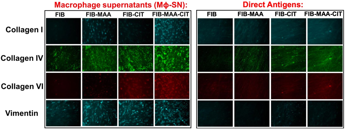
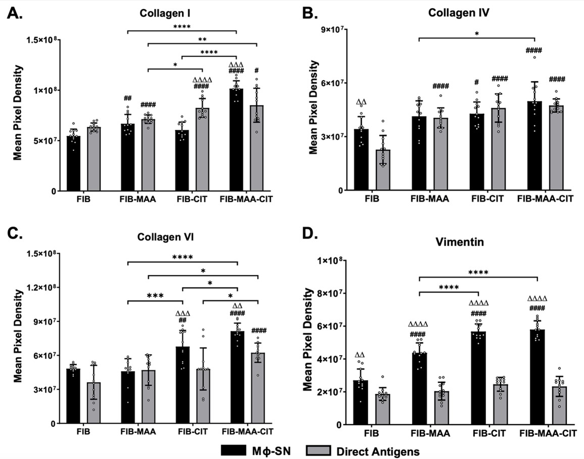
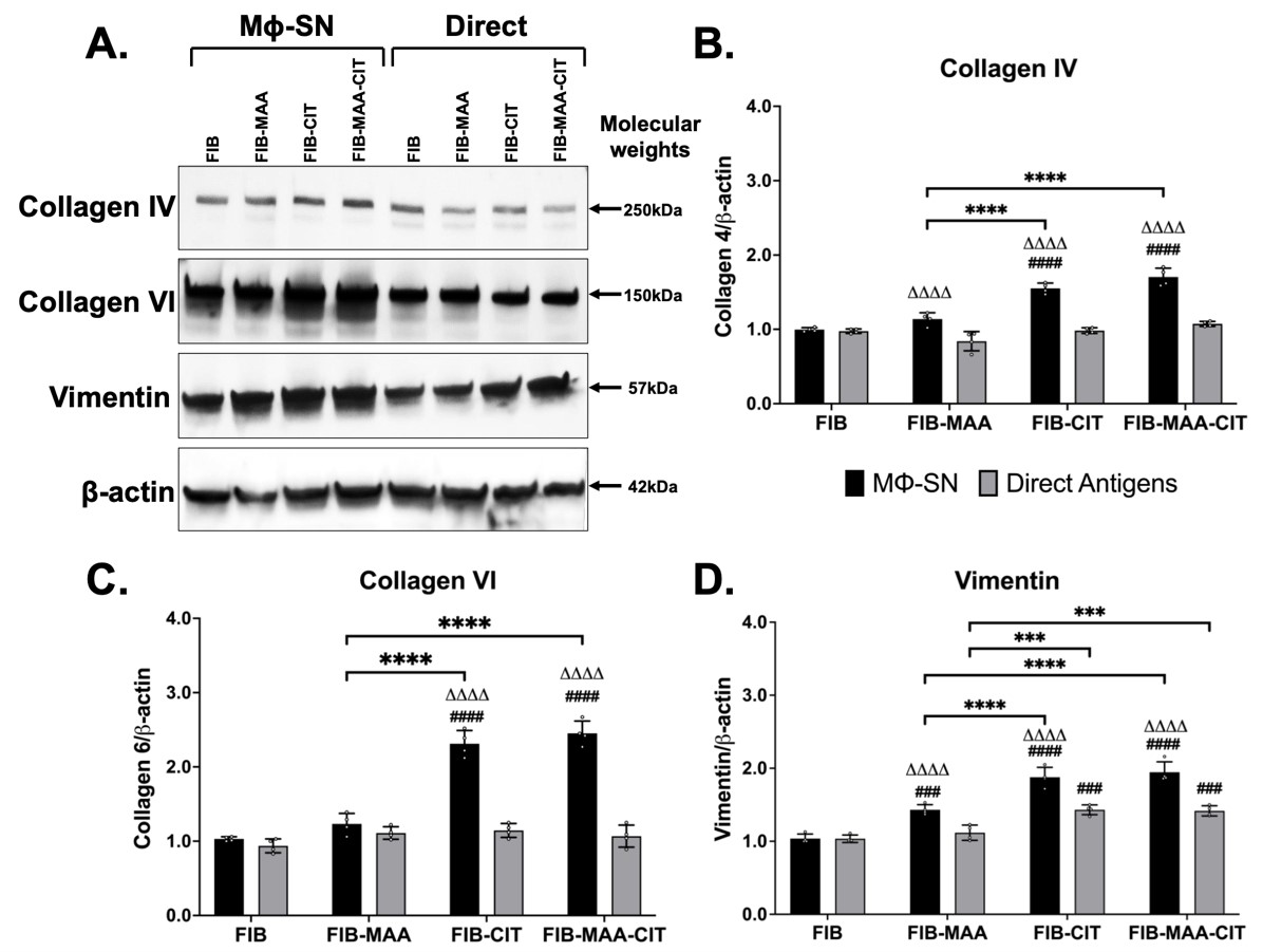
Methods: HLFs were stimulated every 48 hours for 14 days either; directly with FIB, FIB-MAA, FIB-CIT, or FIB-MAA-CIT, or indirectly with supernatants (SN) harvested from activated U937 MΦ (MΦ-SN) stimulated with the same antigens (MΦ-SNFIB-MAA, MΦ-SNFIB-CIT, and MΦ-SNFIB-MAA-CIT). Fluorescent immunohistochemistry (IHC) coupled with mean pixel density evaluations were utilized to monitor morphological changes and to quantify the levels of extracellular matrix (ECM) protein deposition including collagen I, IV, VI, and vimentin. Western blots were used to validate the findings from IHC.

Results: Following antigenic exposure, two phenotypically distinct morphologies were observed in HLF cells. HLF cells stimulated with MΦ-SNFIB-MAA, MΦ-SNFIB-CIT, and MΦ-SNFIB-MAA-CIT demonstrated circular and spindle-shaped changes in cellular morphology consistent with cell activation (Figure 1). HLF cells stimulated with MΦ-SNFIB-MAA-CIT showed the highest deposition of collagen I and VI compared to either single modification (*p< 0.05) or direct antigen stimulation (ΔΔp< 0.01) (Figure 2). By Western Blot, HLFs treated with MΦ-SNFIB-CIT and MΦ-SNFIB-MAA-CIT demonstrated the highest expression of collagen IV, collagen VI, and vimentin compared to MΦ-SNFIB (####p< 0.001), MΦ-SNFIB-MAA (****p< 0.001) or direct antigen stimulation (ΔΔΔΔp< 0.001) (Figure 3).

Conclusion: Findings from this study demonstrate that MΦ exposed to CIT and/or MAA modified FIB secrete soluble factors that activate pro-fibrotic responses in HLF effector cells, that could potentially lead to pulmonary fibrosis associated with RA-ILD. These results suggest that interventions targeting MAA and CIT formation, or cell binding could represent a novel therapeutic approach in the management of RA-ILD.

Supporting image 1

Figure 1: Fluorescent immunohistochemistry (IHC) images of human lung fibroblasts (HLFs) stimulated with macrophage supernatants (MΦ-SN). Following 14-day stimulations (every 48 hours) with MΦ-SN (panel A) or direct antigens (panel B), formalin fixed HLF cells were examined qualitatively for ECM deposition. Cells were stained for collagen (types I, IV, and VI) and vimentin. Compared to direct antigen stimulation (panel B), the morphology of HLFs in MΦ-SN treatment groups (MΦ-SNFIB-MAA, MΦ-SNFIB-CIT, and MΦ-SNFIB-MAA-CIT) shifts to circular/spindle-shape cells (panel A).

Supporting image 2

Figure 2: Mean pixel density quantification of extracellular matrix (ECM) deposition from human lung fibroblasts (HLFs) stimulated with macrophage supernatants (MΦ-SN) using fluorescent immunohistochemistry. ECM deposition was quantified from fluorescent IHC images of formalin fixed HLFs following 14-day stimulations with MΦ-SN or direct antigens. Results shown for minimum of 6 replicate experiments. Staining for collagen types I, IV, VI (panels A-C) and vimentin (panel D) was quantified as mean pixel density. One-way ANOVA with Tukey post-hoc test used to compare staining across treatment groups. Comparisons to MΦ-SNFIB or FIB shown above each bar (####p<0.0001, ##p<0.01, #p<0.05); other significant intergroup comparisons illustrated with horizontal bars (****p<0.0001, ***p<0.001, **p<0.01, *p<0.05). Pairwise differences between MΦ-SN and direct antigen examined using t-test; shown above each bar (ΔΔΔΔp<0.0001, ΔΔΔp<0.001, ΔΔp<0.01).

Supporting image 3

Figure 3: Western blot quantification of extracellular matrix (ECM) deposition from human lung fibroblasts (HLFs) following stimulation with macrophage supernatants (MΦ-SN) or direct antigen. HLFs exposed to MΦ-SN and direct antigen were lysed and examined via Western blot (panel A). Results shown for a minimum of 6 replicate experiments. Quantification for collagen type IV (panel B), collagen type VI (panel C), and vimentin (panel D) shown. Type I collagen not detected (not shown). One-way ANOVA with Tukey post-hoc test used to compare quantities across treatment groups; comparisons to MΦ-SNFIB or FIB illustrated above each bar (####p<0.0001, ###p<0.001); other significant intergroup comparisons shown with horizontal bars (****p<0.0001, ***p<0.001). Pairwise comparisons between MΦ-SN and direct antigen examined using t-test; depicted above each bar (ΔΔΔΔp<0.0001).


Disclosures: L. Klingemann: None; N. Aripova: None; N. Aripova: None; M. Duryee: None; C. Hunter: None; J. Poole: None; B. England: Boehringer-Ingelheim, 5; T. Mikuls: Elsevier, 9, Horizon Therapeutics, 2, 5, Pfizer, 2, Sanofi, 2, UCB Pharma, 2, Wolters Kluwer Health (UpToDate), 9; G. Thiele: None.

To cite this abstract in AMA style:

Klingemann L, Aripova N, Aripova N, Duryee M, Hunter C, Poole J, England B, Mikuls T, Thiele G. Pro-Fibrotic Effects of Malondialdehyde-Acetaldehyde-Adducted and/or Citrullinated Proteins on Macrophages and Human Lung Fibroblasts [abstract]. Arthritis Rheumatol. 2024; 76 (suppl 9). https://acrabstracts.org/abstract/pro-fibrotic-effects-of-malondialdehyde-acetaldehyde-adducted-and-or-citrullinated-proteins-on-macrophages-and-human-lung-fibroblasts/. Accessed .
  • Tweet
  • Click to email a link to a friend (Opens in new window) Email
  • Click to print (Opens in new window) Print

« Back to ACR Convergence 2024

ACR Meeting Abstracts - https://acrabstracts.org/abstract/pro-fibrotic-effects-of-malondialdehyde-acetaldehyde-adducted-and-or-citrullinated-proteins-on-macrophages-and-human-lung-fibroblasts/

Advanced Search

Your Favorites

You can save and print a list of your favorite abstracts during your browser session by clicking the “Favorite” button at the bottom of any abstract. View your favorites »

All abstracts accepted to ACR Convergence are under media embargo once the ACR has notified presenters of their abstract’s acceptance. They may be presented at other meetings or published as manuscripts after this time but should not be discussed in non-scholarly venues or outlets. The following embargo policies are strictly enforced by the ACR.

Accepted abstracts are made available to the public online in advance of the meeting and are published in a special online supplement of our scientific journal, Arthritis & Rheumatology. Information contained in those abstracts may not be released until the abstracts appear online. In an exception to the media embargo, academic institutions, private organizations, and companies with products whose value may be influenced by information contained in an abstract may issue a press release to coincide with the availability of an ACR abstract on the ACR website. However, the ACR continues to require that information that goes beyond that contained in the abstract (e.g., discussion of the abstract done as part of editorial news coverage) is under media embargo until 10:00 AM ET on November 14, 2024. Journalists with access to embargoed information cannot release articles or editorial news coverage before this time. Editorial news coverage is considered original articles/videos developed by employed journalists to report facts, commentary, and subject matter expert quotes in a narrative form using a variety of sources (e.g., research, announcements, press releases, events, etc.).

Violation of this policy may result in the abstract being withdrawn from the meeting and other measures deemed appropriate. Authors are responsible for notifying colleagues, institutions, communications firms, and all other stakeholders related to the development or promotion of the abstract about this policy. If you have questions about the ACR abstract embargo policy, please contact ACR abstracts staff at [email protected].

Wiley

  • Online Journal
  • Privacy Policy
  • Permissions Policies
  • Cookie Preferences

© Copyright 2025 American College of Rheumatology