ACR Meeting Abstracts

ACR Meeting Abstracts

  • Meetings
    • ACR Convergence 2024
    • ACR Convergence 2023
    • 2023 ACR/ARP PRSYM
    • ACR Convergence 2022
    • ACR Convergence 2021
    • ACR Convergence 2020
    • 2020 ACR/ARP PRSYM
    • 2019 ACR/ARP Annual Meeting
    • 2018-2009 Meetings
    • Download Abstracts
  • Keyword Index
  • Advanced Search
  • Your Favorites
    • Favorites
    • Login
    • View and print all favorites
    • Clear all your favorites
  • ACR Meetings

Abstract Number: 18L

Primary CNS Vaculitis Vs Neurosarcoidosis: Can We Predict the Diagnosis with MRI?

Didem Saygin1, Priya Sundaram-Simonelli2, Leonard H. Calabrese3, Elisabeth Ray4, William Messner5, Stephen Jones2, Jinny Tavee6 and Rula A Hajj-Ali7, 1Cleveland Clinic, Cleveland, OH, 2Neuroradiology, Cleveland Clinic, Cleveland, OH, 3Rheumatic & Immunologic Disease, Cleveland Clinic, Cleveland, OH, 4Rheumatology, Cleveland Clinic, Cleveland, OH, 5Quantitative Health Sciences, Cleveland Clinic, Cleveland, OH, 6Neuromuscular diseases, Cleveland Clinic, Cleveland, OH, 7Rheumatic and Immunologic Dis, Cleveland Clinic, Cleveland, OH

Meeting: 2016 ACR/ARHP Annual Meeting

Date of first publication: October 19, 2016

Keywords: central nervous system involvement, Late-Breaking 2016, Magnetic resonance imaging (MRI), neurosarcoidosis and vasculitis

  • Tweet
  • Email
  • Print
Session Information

Date: Tuesday, November 15, 2016

Title: ACR Late-Breaking Poster Session

Session Type: ACR Late-breaking Abstract Session

Session Time: 9:00AM-11:00AM

Background/Purpose:

Neurosarcoidosis and primary angiitis of central nervous system (PACNS) are inflammatory diseases affecting central nervous system, with overlapping clinical and pathological characteristics. Differentiating between both conditions is crucial for differences in therapeutic options. In this study, we aimed to compare the MRI findings between these two conditions to enhance our diagnostic approach, which can lead to more accurate decisions about appropriate treatment strategy.Methods:

Thirty-four patients with PACNS and forty two patients with neurosarcoidosis were included in the study.  Brain and/or spinal cord MRI performed near the time of presentation were blindly evaluated by two neuroradiologists. Data regarding involvement of pachy– and leptomeninges, basal meninges, cranial nerves, cerebral grey and white matter, and spinal cord were recorded for each patient. For each of these sites, presence, pattern (focal vs diffuse), localization (cerebral lobes, brain stem, cerebellum, tentorium, falx cerebri), and laterality of involvement (right, left, bilateral) were recorded. Additionally, ventriculomegaly, mass effect, and parenchymal hemorrhage were recorded. Chi square and Fisher’s exact tests were used to compare the groups, as appropriate. All statistical analyses were performed using SAS 9.4 software.Results:

Mean age of patients was 45.6 (range, 5-80) and 44.1 (range, 25-66) in PACNS and neurosarcoidosis groups, respectively. MRI findings are summarized in the Table. Enhancement and abnormal signal in grey and/or white matter were significantly more common in patients with PACNS. However, localization and laterality of grey and/or white matter involvements did not show a difference between groups. In addition, basal ganglia enhancement and parenchymal hemorrhage were significantly associated with PACNS. On the other hand, patients with neurosarcoidosis had more common basal meningeal, spinal cord and cranial nerve involvements. Frequency, pattern, laterality and localization of pachy– and leptomeningeal, pituitary and hypothalamic involvements; frequency of brain mass effect and ventriculomegaly were similar between groups.Conclusion:

Patients with PACNS had higher frequency of cerebral and basal ganglia enhancement, and parenchymal hemorrhage, while patients with neurosarcoidosis demonstrated more frequent spinal cord, basal meningeal and cranial nerve involvements. These findings suggest that MRI can be an efficient tool in distinguishing PACNS from neurosarcoidosis. Table. Frequencies and comparison of MRI findings in patients with PACNS and neurosarcoidosis (NS).  


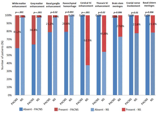
Disclosure: D. Saygin, None; P. Sundaram-Simonelli, None; L. H. Calabrese, None; E. Ray, None; W. Messner, None; S. Jones, None; J. Tavee, None; R. A. Hajj-Ali, None.

To cite this abstract in AMA style:

Saygin D, Sundaram-Simonelli P, Calabrese LH, Ray E, Messner W, Jones S, Tavee J, Hajj-Ali RA. Primary CNS Vaculitis Vs Neurosarcoidosis: Can We Predict the Diagnosis with MRI? [abstract]. Arthritis Rheumatol. 2016; 68 (suppl 10). https://acrabstracts.org/abstract/primary-cns-vaculitis-vs-neurosarcoidosis-can-we-predict-the-diagnosis-with-mri/. Accessed .
  • Tweet
  • Email
  • Print

« Back to 2016 ACR/ARHP Annual Meeting

ACR Meeting Abstracts - https://acrabstracts.org/abstract/primary-cns-vaculitis-vs-neurosarcoidosis-can-we-predict-the-diagnosis-with-mri/

Advanced Search

Your Favorites

You can save and print a list of your favorite abstracts during your browser session by clicking the “Favorite” button at the bottom of any abstract. View your favorites »

All abstracts accepted to ACR Convergence are under media embargo once the ACR has notified presenters of their abstract’s acceptance. They may be presented at other meetings or published as manuscripts after this time but should not be discussed in non-scholarly venues or outlets. The following embargo policies are strictly enforced by the ACR.

Accepted abstracts are made available to the public online in advance of the meeting and are published in a special online supplement of our scientific journal, Arthritis & Rheumatology. Information contained in those abstracts may not be released until the abstracts appear online. In an exception to the media embargo, academic institutions, private organizations, and companies with products whose value may be influenced by information contained in an abstract may issue a press release to coincide with the availability of an ACR abstract on the ACR website. However, the ACR continues to require that information that goes beyond that contained in the abstract (e.g., discussion of the abstract done as part of editorial news coverage) is under media embargo until 10:00 AM ET on November 14, 2024. Journalists with access to embargoed information cannot release articles or editorial news coverage before this time. Editorial news coverage is considered original articles/videos developed by employed journalists to report facts, commentary, and subject matter expert quotes in a narrative form using a variety of sources (e.g., research, announcements, press releases, events, etc.).

Violation of this policy may result in the abstract being withdrawn from the meeting and other measures deemed appropriate. Authors are responsible for notifying colleagues, institutions, communications firms, and all other stakeholders related to the development or promotion of the abstract about this policy. If you have questions about the ACR abstract embargo policy, please contact ACR abstracts staff at [email protected].

Wiley

  • Online Journal
  • Privacy Policy
  • Permissions Policies
  • Cookie Preferences

© Copyright 2025 American College of Rheumatology