Session Information
Session Type: ACR Poster Session A
Session Time: 9:00AM-11:00AM
Background/Purpose: Positron emission tomography (PET)-CT scan can assess large vessel vasculitis in giant cell arteritis (GCA). It has a reported sensitivity of 80% for biopsy positive disease but the superficial temporal, occipital and vertebral arteries have not previously been assessed due to difficulties with spatial resolution.
Methods: 41 patients suspected of having GCA between May 2016 and April 2017 underwent fluorine-18 fluoro-2-deoxyglucose (FDG) time-of-flight PET-CT scan within 72 hours of commencing corticosteroids and before clinically guided temporal artery biopsy (TAB). Patients were imaged from the vertex of the head to the diaphragm with 1mm CT slice reconstruction. FDG uptake was graded at 16 vascular sites using a semi-quantitative scale defined as zero (less than or equal to blood pool), one (minimally increased), two (clearly increased) or three (very marked uptake) by one of two blinded, experienced nuclear medicine physicians. Arterial calcification, shoulder region FDG uptake, infection and cancer were also reported.
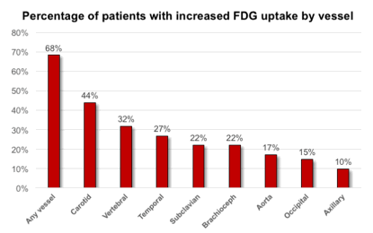
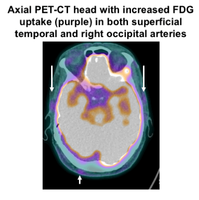
Results: Mean age was 70 and 29 (71%) were female. Headache (88%), jaw claudication (34%), PMR (32%) and visual disturbance (29%) were common reported symptoms. Median CRP was 16 mg/L (range 1 – 299) and ESR was 36 mm/hr (range 5 – 130). Eight (20%) patients had mural inflammation on biopsy, four (10%) had limited periadventitial small vessel vasculitis and one had unequivocal thoracic aortitis on CT scan and did not undergo TAB. 28 (68%) had negative biopsies. 30 (73%) met the 1990 ACR criteria for GCA and 18 (44%) were assessed by the treating clinician as having definite or probable (>= 50% chance) GCA at two-week follow-up. 28 (68%) had at least grade one uptake in one or more vascular territory and 14 (34%) had at least grade two uptake. The carotid (44%), vertebral (32%) and superficial temporal (27%) arteries were the most affected. 6 (15%) patients had increased uptake isolated to the vertebral, temporal or occipital arteries. Shoulder region uptake was detected in 23 (56%) of patients, infection in three (two sinusitis, one pneumonia) and lung cancer in three. Vascular calcification did not globally correlate with increased FDG uptake (Spearman’s correlation coefficient = 0.05).
Conclusion: 68% of patients initially suspected of having GCA had at least mild increased FDG uptake on PET-CT scan. This compared with only 30% with inflammatory changes on biopsy. Increased uptake was commonly seen in the vertebral and temporal arteries which have not previously been assessed in PET-CT studies. Long-term follow-up will determine the clinical role of this protocol in GCA.
To cite this abstract in AMA style:
Sammel A, Hsiao E, Nguyen K, Schembri G, Schrieber L, Youssef P, Janssen B, Laurent R. Prevalence and Distribution of Vascular FDG Uptake on Positron Emission Tomography (PET)-CT in Patients Suspected of Having Giant Cell Arteritis – Interim Results from the Giant Cell Arteritis and PET Scan (GAPS) Study [abstract]. Arthritis Rheumatol. 2017; 69 (suppl 10). https://acrabstracts.org/abstract/prevalence-and-distribution-of-vascular-fdg-uptake-on-positron-emission-tomography-pet-ct-in-patients-suspected-of-having-giant-cell-arteritis-interim-results-from-the-giant-cell-arteritis/. Accessed .« Back to 2017 ACR/ARHP Annual Meeting
ACR Meeting Abstracts - https://acrabstracts.org/abstract/prevalence-and-distribution-of-vascular-fdg-uptake-on-positron-emission-tomography-pet-ct-in-patients-suspected-of-having-giant-cell-arteritis-interim-results-from-the-giant-cell-arteritis/


