Session Information
Session Type: Abstract Submissions (ACR)
Background/Purpose:
Abatacept inhibits the costimulatory interaction of T-lymphocytes and antigen-presenting cells. Treatment of early and active primary Sjögren’s Syndrome (pSS) with abatacept decreases disease activity. The aim of this study was to assess the histopathological changes in the parotid gland tissue after treatment with abatacept in pSS patients.
Methods:
15 patients (12 female, 3 male) were included in the open-label Active Sjögren Abatacept Pilot (ASAP) study and received 8 intravenous abatacept infusions on days 1,15, 29 and every 4 weeks thereafter. Before treatment and at 25 weeks of follow up a parotid gland biopsy was taken. Hematoxilin-eosin stains were evaluated for focus score (foci of 50 lymphocytes/4mm2), lymphoepithelial lesions (LELs) and presence of germinal centers. A CD45 stain was used to calculate the area of lymphocytic infiltrate (Aperio ImageScope v12.0). The infiltrate was further analysed for numbers of CD20+ B-cells, CD3+ T-cells and IgA, IgG and IgM positive plasma cells using HistoQuest.
Results:
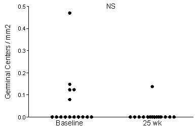
At baseline 5 out of 15 patients (33%) showed presence of germinal centers in parotid gland tissue. In all these 5 patients germinal centers were absent after abatacept treatment. One patient showed only limited germinal center activity after treatment. The mean number of germinal centers decreased from 0,06 GC/mm2 to 0,01 GC/mm2. Importantly, the number of germinal centers/mm2 at baseline is associated with a clinical improvement in the glandular domain of the ESSDAI. Abatacept treatment did not affect focusscore, LELs, amount of infiltrated B-cells and T-cells. Analysis of the plasma cell population is currently in progress.
Figure 1: Number of germinal centers in parotid gland tissue decline after treatment with abatacept.
Conclusion:
Presence of germinal centers at baseline in parotid gland tissue is associated with clinical respons in the glandular domain of the ESSDAI after treatment with abatacept in primary Sjogren’s syndrome. Histopathological analysis of parotid gland tissue may therefore be helpfull in assessing treatment efficacy. As expected, abatacept does not affect the lymphocytic infiltrate in terms of overall numbers of T- and B-cells.
Disclosure:
E. A. Haacke,
None;
F. G. M. Kroese,
None;
P. M. Meiners,
None;
B. van der Vegt,
None;
A. Vissink,
None;
F. K. L. Spijkervet,
None;
H. Bootsma,
BMS,
2.
« Back to 2014 ACR/ARHP Annual Meeting
ACR Meeting Abstracts - https://acrabstracts.org/abstract/presence-of-germinal-centers-at-baseline-is-associated-with-clinical-response-of-glandular-essdai-domain-after-abatacept-treatment-in-primary-sjogrens-syndrome/

