Session Information
Session Type: Abstract Submissions (ACR)
Background/Purpose: Rheumatoid arthritis is a chronic inflammatory disease manifested by episodic flares in affected joints that are challenging to predict and treat. Longitudinal contrast enhanced-MRI (CE-MRI) of inflammatory arthritis in tumor necrosis factor-transgenic (TNF-Tg) mice has demonstrated that popliteal lymph nodes (PLN) increase in volume and contrast enhancement during the pre-arthritic “expanding” phase, and then suddenly “collapse” during knee flare. Given the potential of this biomarker of arthritic flare, we aimed to develop a cost-effective means of phenotyping PLN using ultrasound (US) imaging.
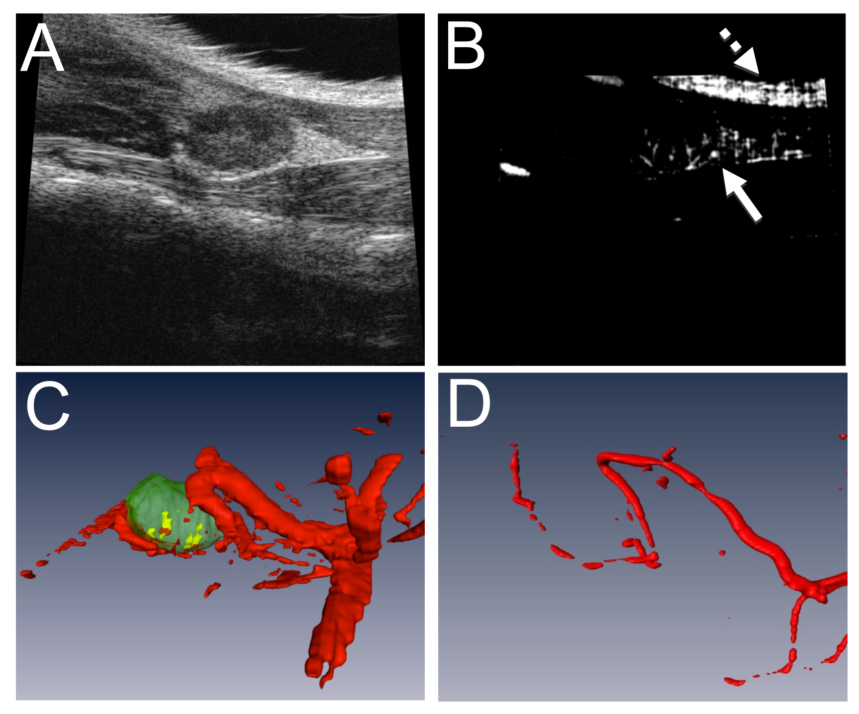
Methods: Power Doppler (PD) US was performed on TNF-Tg mice whose PLN were previously phenotyped as expanding or collapsed using CE-MRI. B-mode (Figure 1A) and PD (Figure 1B) 3D scans were used to generate 3D volume estimates (Figure 1C) to calculate the PD volume normalized to the PLN volume, or the normalized power Doppler volume (NPDV). To verify this methodology for vessels near the PLN, a mouse underwent Microfil perfusion and micro-CT to confirm the US structures were blood vessels near the PLN (Figure 1D).
Results: PD-US demonstrates that expanding PLN have a significantly higher NPDV versus collapsed PLN (0.553±0.007 vs. 0.008±0.003 mm3; p<0.05). Moreover, we define the upper (>0.030 mm3) and lower (<0.016 mm3) quartile NPDVs in this cohort of mice, which serve as conservative thresholds to phenotype PLN as expanding and collapsed, respectively. To observe agreement between PD-US and CE-MRI, PLN were phenotyped as collapsed by PD-US and then underwent CE-MRI. Of the 12 PLN in the study, there was disagreement in 4 cases. However, these 4 cases presented with marked synovitis in the adjacent knee, a hallmark of arthritic flare, which occurs after PLN collapse. Therefore, we find that PD-US correctly phenotyped all of the PLN in this cohort.
Conclusion: We find PD-US to be more accurate for phenotyping PLN than CE-MRI. PD-US permits the PLN in the middle quartiles to be subsequently phenotyped with longitudinal scans and studied accordingly. This is a major advantage over CE-MRI phenotyping, which is too costly to utilize for this purpose. PD-US is both a safe and feasible approach and can be used to phenotype PLN as expanding versus collapsed for prospective studies.
Figure 1. Quantification of NPDV using PD-US and validation with vascular micro-CT. PD-US was performed to generate the 2D images obtained using B mode (A) and PD (B) scans of the PLN. Note the artifact in the PD due to the surface of the skin (dashed arrow in B) versus the true PD signal (solid arrow in B). The PLN (green structure in C) was used to apply a mask to the total blood volume (red structures in C) to give the total PD volume within the PLN (yellow structure in C). To confirm this methodology, the mouse underwent Microfil perfusion and the micro-CT is shown (D). Note the similarities in vessel structure with decreased vessel diameter due to live in vivo imaging vs. perfusion.
Disclosure:
E. M. Bouta,
None;
Y. Ju,
None;
H. Rahimi,
None;
R. Wood,
None;
L. Xing,
None;
E. M. Schwarz,
None.
« Back to 2013 ACR/ARHP Annual Meeting
ACR Meeting Abstracts - https://acrabstracts.org/abstract/power-doppler-ultrasound-phenotyping-of-expanding-versus-collapsed-popliteal-lymph-nodes-in-murine-inflammatory-arthritis/

