Session Information
Date: Sunday, November 7, 2021
Session Type: Poster Session B
Session Time: 8:30AM-10:30AM
Background/Purpose: In previous studies vertebral corner inflammation (VCI) and vertebral corner fat deposition (VCFD) were associated with syndesmophyte formation on cervical and lumbar conventional radiography. We studied associations between patterns of VCI, VCFD and a combination of both on magnetic resonance imaging (MRI) and the development of new or grown syndesmophytes on whole spine low dose computed tomography (ldCT), thereby studying these associations also in the thoracic spine.
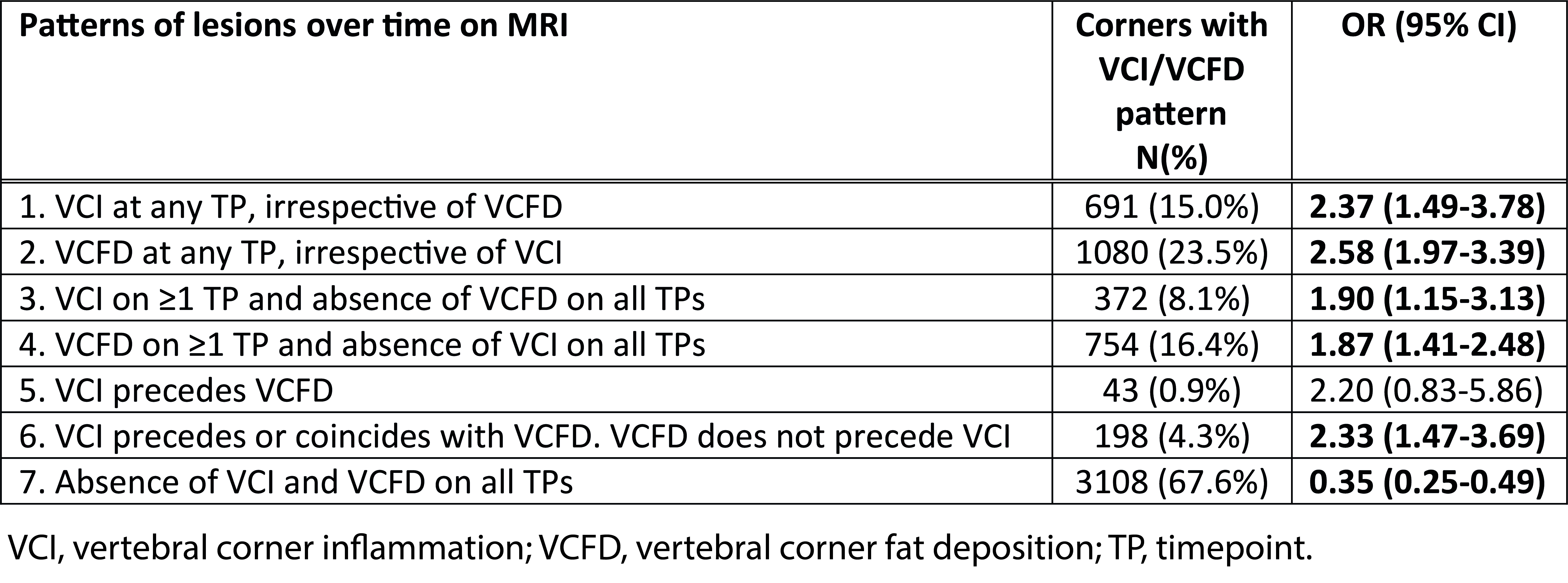
Methods: Patients in the Sensitive Imaging in Ankylosing Spondylitis cohort underwent MRI at baseline, 1 year and 2 years, and ldCT at baseline and 2 years. MRI lesions were scored by 3 central readers, using the SPARCC method for VCI and the CanDen method for VCFD, and coded as absent or present per timepoint and per reader. MRI patterns over time (Table) were deemed present if seen by ≥2 out of 3 readers. The patterns reflect hypothetical associations between presence and absence of VCI and VCFD, independently and combined, on ldCT detected new or grown syndesmophytes. Individual reader change scores were used for ldCT images, scored by 2 central readers with the Computed Tomography Syndesmophyte Score. New (CTSS 0 à 1, 2 or 3) and grown (CTSS 1 à 2 or 3; 2 à 3) syndesmophytes were grouped together to represent bone formation. Corners not at risk for the outcome due to presence of a bridged syndesmophyte at baseline were excluded. Multilevel generalized estimated equations were used, with separate models per MRI pattern, accounting for correlations within patients and between ldCT readers.
Results: Fifty patients were included, contributing a total of 4600 vertebral corners. Their mean age was 49.3 years (SD 9.8), 86% were male and 78% were HLA-B27+. Presence of VCI and VCFD patterns ranged from 43 (0.9%) to 3108 (67.6%) corners (Table), with the lowest frequency being for VCI preceding VCFD. Protection against syndesmophyte development was seen in case of absence of both VCI and VCFD (OR 0.35) and positive associations with ORs ranging from 1.87-2.58 were observed for various VCI/VCFD patterns. Nevertheless, out of all corners with a new or grown syndesmophyte, 47.3% of corners according to reader 1 and 43.9% according to reader 2 had neither VCI nor VCFD preceding the bone formation.
Conclusion: Presence of VCI or VCFD and combinations of the two increased odds of bone formation 2 years later, whereas absence of both VCI and VCFD decreased the odds, showing that VCI and VCFD have some role in the development of syndesmophytes. However, almost half of all bone formation occurred in corners without VCI or VCFD, suggesting the presence of these lesions in yearly MRIs does not fully explain the development of syndesmophytes. This study confirmed that there is an association between VCI and VCFD and bone formation also for the thoracic spine and on ldCT compared to conventional radiography.
To cite this abstract in AMA style:
Stal R, Baraliakos X, Sepriano A, van Gaalen F, Ramiro S, van den Berg R, Reijnierse M, Braun J, Landewé R, van der Heijde D. MRI Vertebral Corner Inflammation and Fat Deposition Are Associated with Whole Spine Low Dose CT Detected Syndesmophytes: A Multilevel Analysis [abstract]. Arthritis Rheumatol. 2021; 73 (suppl 9). https://acrabstracts.org/abstract/mri-vertebral-corner-inflammation-and-fat-deposition-are-associated-with-whole-spine-low-dose-ct-detected-syndesmophytes-a-multilevel-analysis/. Accessed .« Back to ACR Convergence 2021
ACR Meeting Abstracts - https://acrabstracts.org/abstract/mri-vertebral-corner-inflammation-and-fat-deposition-are-associated-with-whole-spine-low-dose-ct-detected-syndesmophytes-a-multilevel-analysis/