ACR Meeting Abstracts

ACR Meeting Abstracts

  • Meetings
    • ACR Convergence 2024
    • ACR Convergence 2023
    • 2023 ACR/ARP PRSYM
    • ACR Convergence 2022
    • ACR Convergence 2021
    • ACR Convergence 2020
    • 2020 ACR/ARP PRSYM
    • 2019 ACR/ARP Annual Meeting
    • 2018-2009 Meetings
    • Download Abstracts
  • Keyword Index
  • Advanced Search
  • Your Favorites
    • Favorites
    • Login
    • View and print all favorites
    • Clear all your favorites
  • ACR Meetings

Abstract Number: 1810

Modulation of Type I Interferon Signaling by Anifrolumab Alters the Spatial Immune Landscape in Cutaneous Lupus Erythematosus

Ksenia Anufrieva1, Rochelle Castillo2, Ce Gao2, Jessica Liu2, Neda Shahriari2, Kimberly Hashemi2, Dustin Taylor3, Rachael Rowley2, Elizabeth Rainone2, Avery LaChance2, Rachel Gate4, ilya Korsunsky2, Ruth Ann Vleugels5 and Kevin Wei6, 1BWH, Cambridge, MA, 2Brigham and Women's Hospital, Boston, MA, 3Vanderbilt University Medical Center, Nashville, TN, 410x Genomics, Pleasanton, CA, 5Brigham and Women's Hospital, Brookline, MA, 6Brigham and Women's Hospital at Harvard Medical School, Boston, MA

Meeting: ACR Convergence 2024

Keywords: Gene Expression, genomics, Inflammation, Systemic lupus erythematosus (SLE), Therapy, alternative

  • Tweet
  • Click to email a link to a friend (Opens in new window) Email
  • Click to print (Opens in new window) Print
Session Information

Date: Monday, November 18, 2024

Title: SLE – Etiology & Pathogenesis Poster

Session Type: Poster Session C

Session Time: 10:30AM-12:30PM

Background/Purpose: Reliable and effective treatments for refractory cutaneous lupus erythematosus (CLE) remained elusive until the arrival of anifrolumab, a monoclonal antibody targeting the type I interferon receptor subunit 1. In this study, we conducted high-resolution spatial and single-cell analysis from paired lesional and non-lesional skin samples of CLE patients pre- and post-anifrolumab treatment to gain insights into the immunopathogenesis of CLE and anifrolumab-induced changes in the cellular milieu. Our initial investigation highlighted significant changes in the spatial organization of immune cells, particularly in regions with increased expression of genes activated by type I interferon.

Methods: Punch biopsies of lesional and non-lesional skin from patients with refractory CLE were obtained prior to and three months after treatment with anifrolumab. We analyzed these formalin-fixed, paraffin-embedded (FFPE) skin biopsies using a single-cell gene expression method (Chromium Single Cell Gene Expression Flex) and a high-resolution spatial transcriptomics technique (Xenium In Situ), sequencing 27 and 17 skin biopsies respectively. This comprehensive approach facilitated the identification and spatial characterization of 53 different cell types.

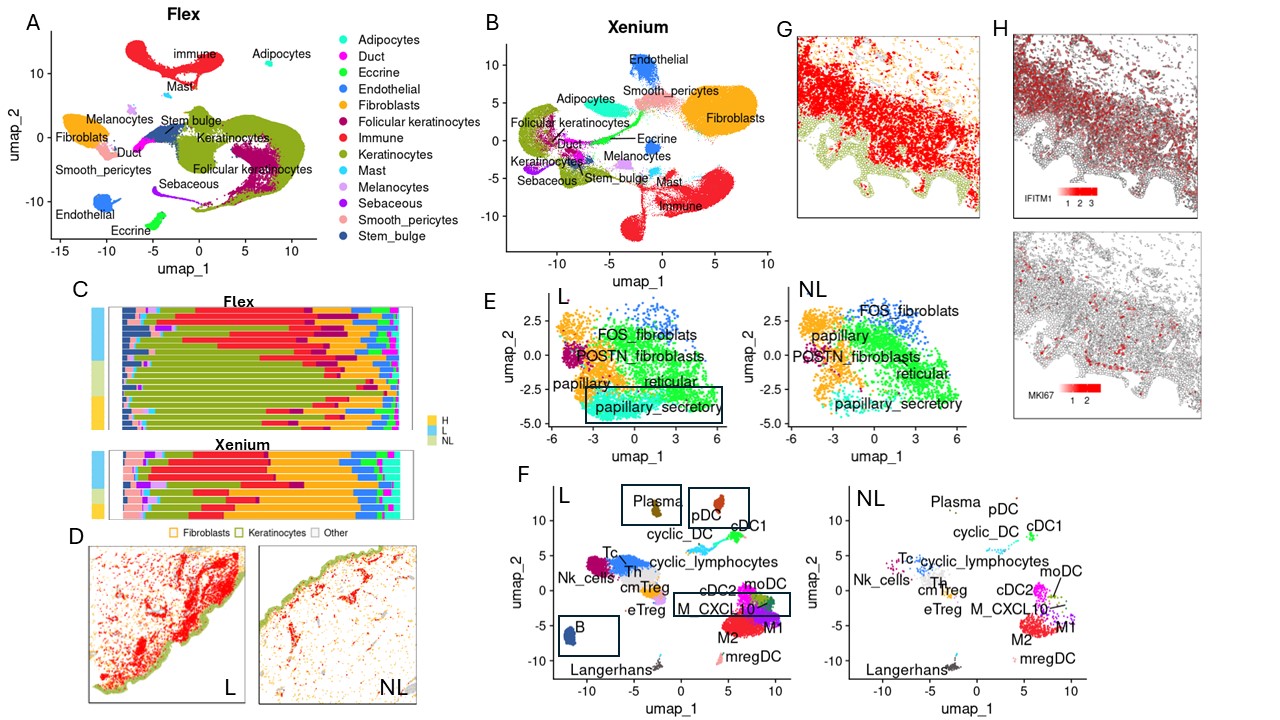
Results: Comparing single cell data from lesional and non-lesional skin biopsies revealed a significant increase in immune cell infiltration, which correlated with elevated levels of cells exhibiting a type I interferon response and proliferative phenotypes (Fig1, A-H). Additionally, pro-inflammatory clusters, including CXCL10-expressing macrophages and cytokine-enriched papillary fibroblasts, emerged in lesional skin (Fig1, E-F). Cell composition analysis demonstrated prevalence of plasmacytoid dendritic cells (pDCs), B cells, plasma cells, and CXCL10+ macrophages exclusively in lesional skin (Fig1, E-F). Spatial mapping across 17 skin biopsies unveiled two distinct clusters of immune aggregates in CLE lesional skin: one highly enriched mregDCs and pDCs occurring near basal keratinocytes, and another characterized by a predominance of macrophages located perivascularly (Fig2, A). Furthermore, analysis of spatial and single cell data from patients treated with anifrolumab demonstrated that reduced type I interferon signaling led to the disappearance of disease-associated clusters and depletion of spatially-organized immune aggregates (Fig2, B-D).

Conclusion: Inhibition of type I interferon signaling induces a shift of the lesional skin transcriptome towards the non-lesional phenotype, characterized by resolution of pro-inflammatory clusters and spatially-distinct immune aggregates. Notably, despite clinical improvement, treatment with anifrolumab did not result in the conversion of either the lesional or non-lesional skin transcriptome into the healthy phenotype. Further research is needed to elucidate the specific molecular response mechanisms underlying the superiority of anifrolumab over other therapies targeting dysregulated interferon signaling.

Supporting image 1

(A) Uniform Manifold Approximation and Projection (UMAP) plot of cells from 27 skin biopsies, identified by single cell FFPE sequencing (scFFPE-seq). Sample composition: 9 lesional untreated (L), 6 healthy (H), 6 non-lesional untreated (NL), 3 lesional treated (TL), and 3 non-lesional treated (TNL). (B) UMAP plot of cells from 17 skin biopsies, identified by Xenium, clustered into 13 major cell types. Sample composition: 5 lesional untreated (L), 2 healthy (H), 3 non-lesional untreated (NL), 4 lesional treated (TL), and 3 non-lesional treated (TNL). (C) Proportion of each cell type in skin samples sequenced by scFFPE-seq and Xenium. (D) Cell type map from Xenium data. Colors: green – keratinocytes, yellow – fibroblasts, red – immune cells, grey – other cells. (E-F) UMAP analysis of fibroblast and immune cell transcriptomes in lesional (L) and non-lesional (NL) skin. (G) Cell type map from Xenium data in lesional skin. Colors: green – keratinocytes, yellow – fibroblasts, red – immune cells, grey – other cells. (H) Xenium spatial plot showing normalized expression of interferon-response marker IFITM1 and proliferative marker MKI67 in lesional skin.

Supporting image 2

(A) Cell type map from Xenium data in lesional skin with two representative immune niches.
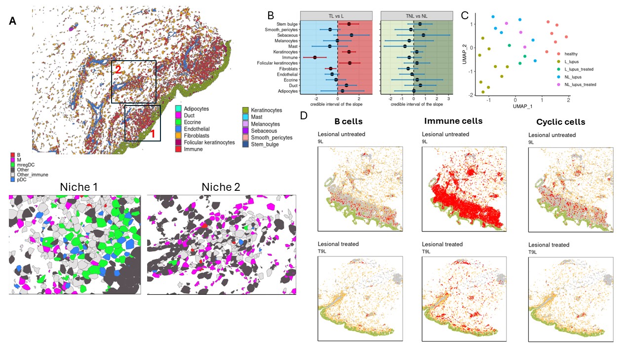
(B) A plot of estimates of differential composition analysis for major cell types between treated and untreated lesional and non-lesional skin samples. The error bars represent 95% confidence intervals. The error bar is blue if the composition of the target cell type is insignificant and red if significant. (C) UMAP of Multicellular Factor Analysis factor scores showing the variability of all cell types captured by single cell Flex sequencing. (D) Cell type map from Xenium data in lesional skin before and after treatment. Colors: green – keratinocytes, yellow – fibroblasts, red – B cells/immune cells/cells with a proliferative phenotype.


Disclosures: K. Anufrieva: None; R. Castillo: None; C. Gao: None; J. Liu: None; N. Shahriari: None; K. Hashemi: None; D. Taylor: None; R. Rowley: None; E. Rainone: None; A. LaChance: GuidePoint, 2, Janssen, 1, Merck, 5, Pfizer, 5, TD Cowen, 2, UCB, 1; R. Gate: 10x Genomics, 3; i. Korsunsky: Mestag Therapeutics LTD, 2; R. Vleugels: Argenx, 2, AstraZeneca, 2, Pfizer, 12, Study PI, Priovant, 2; K. Wei: 10X Genomics, 5, Gilead sciences, 2, Merck/MSD, 5, Mestag, 2, santa ana bio, 2.

To cite this abstract in AMA style:

Anufrieva K, Castillo R, Gao C, Liu J, Shahriari N, Hashemi K, Taylor D, Rowley R, Rainone E, LaChance A, Gate R, Korsunsky i, Vleugels R, Wei K. Modulation of Type I Interferon Signaling by Anifrolumab Alters the Spatial Immune Landscape in Cutaneous Lupus Erythematosus [abstract]. Arthritis Rheumatol. 2024; 76 (suppl 9). https://acrabstracts.org/abstract/modulation-of-type-i-interferon-signaling-by-anifrolumab-alters-the-spatial-immune-landscape-in-cutaneous-lupus-erythematosus/. Accessed .
  • Tweet
  • Click to email a link to a friend (Opens in new window) Email
  • Click to print (Opens in new window) Print

« Back to ACR Convergence 2024

ACR Meeting Abstracts - https://acrabstracts.org/abstract/modulation-of-type-i-interferon-signaling-by-anifrolumab-alters-the-spatial-immune-landscape-in-cutaneous-lupus-erythematosus/

Advanced Search

Your Favorites

You can save and print a list of your favorite abstracts during your browser session by clicking the “Favorite” button at the bottom of any abstract. View your favorites »

All abstracts accepted to ACR Convergence are under media embargo once the ACR has notified presenters of their abstract’s acceptance. They may be presented at other meetings or published as manuscripts after this time but should not be discussed in non-scholarly venues or outlets. The following embargo policies are strictly enforced by the ACR.

Accepted abstracts are made available to the public online in advance of the meeting and are published in a special online supplement of our scientific journal, Arthritis & Rheumatology. Information contained in those abstracts may not be released until the abstracts appear online. In an exception to the media embargo, academic institutions, private organizations, and companies with products whose value may be influenced by information contained in an abstract may issue a press release to coincide with the availability of an ACR abstract on the ACR website. However, the ACR continues to require that information that goes beyond that contained in the abstract (e.g., discussion of the abstract done as part of editorial news coverage) is under media embargo until 10:00 AM ET on November 14, 2024. Journalists with access to embargoed information cannot release articles or editorial news coverage before this time. Editorial news coverage is considered original articles/videos developed by employed journalists to report facts, commentary, and subject matter expert quotes in a narrative form using a variety of sources (e.g., research, announcements, press releases, events, etc.).

Violation of this policy may result in the abstract being withdrawn from the meeting and other measures deemed appropriate. Authors are responsible for notifying colleagues, institutions, communications firms, and all other stakeholders related to the development or promotion of the abstract about this policy. If you have questions about the ACR abstract embargo policy, please contact ACR abstracts staff at [email protected].

Wiley

  • Online Journal
  • Privacy Policy
  • Permissions Policies
  • Cookie Preferences

© Copyright 2025 American College of Rheumatology