
Session Information
Session Type: Abstract Submissions (ACR)
Background/Purpose: Knee joint replacement (KR) is a cost-effective procedure with good long-term outcomes. However, there is no clear consensus on indications for KR. Subchondral bone marrow lesions (BMLs) have been identified as important structural features relevant to clinical manifestation (e.g.pain) as well as structural progression (e.g. cartilage loss). Thus, BMLs are promising biomarkers for structural progression to important clinical outcomes such as KR. The aims of this study were therefore to test whether presence and size of BMLs increased odds of KR, and whether worsening of BMLs over time increased odds of KR.
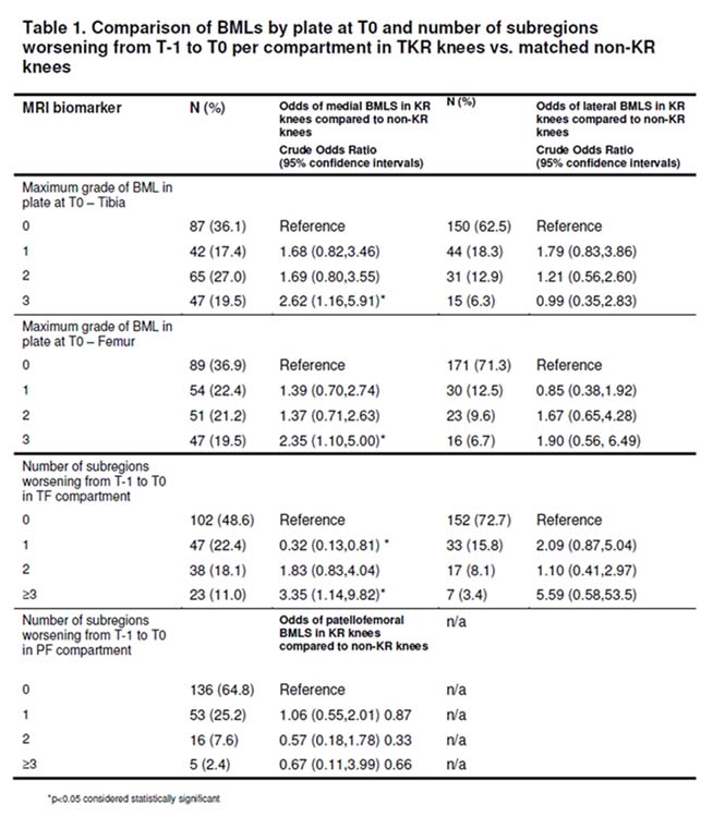
Methods: We studied 121 knees from OAI participants that underwent KR before the 48 month visit at two time points (T0 and T-1) prior to KR, (i.e. for a KR reported at the 48 month (M) visit, T0 = 36M and T-1 = 24M). These were matched with 121 control knees that did not undergo KR based on radiographic disease stage, sex, and age (+/- 5y). 3Tesla MRIs were read for subchondral BMLs in 14 articular subregions using the semiquantitative MOAKS system. Only BML size, which is scored from 0-3, was considered in this study. Analyses were performed on a plate (medial tibia, medial femur, lateral tibia, lateral femur, trochlea, patella) and compartmental level (medial tibio-femoral joint [TFJ], lateral TFJ and patello-femoral joint – [PFJ]). Conditional logistic regression was applied to assess the risk of KR relative to the maximum BML size per plate at T0. In addition, the number of subregions per compartment showing BML worsening from the time point prior T0 (=T-1) to T0 was analyzed relative to the odds of KR following T0.
Results: Subjects were on average 65.3 years old (SD ± 8.6), predominantly female (58.1%) and overweight (mean BMI 29.6 SD ± 4.9). The odds of KR were significantly greater for knees exhibiting large (i.e. grade 3) BMLs in the medial compartment when compared to the knees without BMLs at T0 (Table 1). Further, the odds for KR were significantly greater for knees with ≥3 subregions exhibiting increase in BML size in the medial TF compartment from T-1 to T0 compared to knees with no subregions showing worsening (OR 3.35, 95%CI 1.14-9.82). No significant associations were found for smaller lesions, the lateral TFJ and PFJ and respective plates cross-sectionally or longitudinally.
Conclusion: On a plate-level analysis, presence of large BMLs in the medial femur and/or tibia at the time point prior to KR was associated with increased odds of KR, while presence of large BMLs in the lateral TFJ or in the PFJ was not. Worsening of BML size in ≥3 subregions in the medial TF compartment from T-1 to T0 was associated with increased odds of KR when compared to knees without worsening in any subregion in the same compartment.
Disclosure:
F. Roemer,
Boston Imaging Core Lab,
1,
National Institute of Health,
5,
Merck Serono,
5;
C. K. Kwoh,
Novartis Pharmaceutical Corporation,
5;
D. Hunter,
Australia Research Council Future Fellowship,
2,
DonJoy,
5,
NIH,
5,
Stryker,
5;
M. J. Hannon,
None;
R. M. Boudreau,
None;
F. Eckstein,
Chondrometrics GmbH,
3,
Chondrometrics GmbH,
4,
Novartis AG,
2,
Novartis, MerckSeronoSanofi Aventis, Abbot, Perceptive, Bioclinica,
5;
Z. Wang,
None;
M. R. John,
Novartis Pharma AG,
1,
Novartis Pharma AG,
3;
A. Guermazi,
Boston Imaging Core Lab,
1,
Stryker,
5,
Merck Serono,
5,
Genzyme Corporation,
5,
AstraZeneca,
5,
Novartis Pharmaceutical Corporation,
5.
« Back to 2012 ACR/ARHP Annual Meeting
ACR Meeting Abstracts - https://acrabstracts.org/abstract/medial-subchondral-bone-marrow-lesions-increase-the-odds-of-knee-joint-replacement-data-from-the-osteoarthritis-initative/