Session Information
Date: Tuesday, November 10, 2015
Title: Vasculitis III
Session Type: ACR Concurrent Abstract Session
Session Time: 2:30PM-4:00PM
Background/Purpose: The
arterial wall is nourished by microvessels (vasa vasorum), which are a potential route of entry for
inflammatory cells in vasculitis. Little is known
about the anatomy of temporal artery vasa vasorum or their relationship to the inflammatory
neovessels in giant cell arteritis.
Methods: Temporal
arteries from consenting patients were serially sectioned (5µm) and stained with haematoxylin and
eosin (H&E). Slides were scanned at 0.5µm/pixel generating ‘virtual copies’. Virtual slides were
registered (aligned) and segmented (annotated). Area and circularity
(isoperimetric quotient) were calculated for microvessels
(vasa vasorum and neovessels).
2D segmentations were connected throughout the virtual case using Medical Image
Manager (Heterogenius Ltd, Leeds), creating 3D
iso-surfaced images.
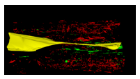
Results: Vasa
vasorum (red) formed a dense plexus within the adventitia of the normal
temporal artery (lumen in yellow), becoming smaller and more numerous towards
the internal elastic lamina. In a completely-occluded
temporal artery with panarteritis, microvessels penetrated all layers of the vascular wall.
Leukocyte aggregates surrounded some of the adventitial vasa vasorum. In a
temporal artery with partial luminal occlusion, the segment with greater
occlusion had a greater number of neovessels (innermost neovessels shown in
green). In GCA, median microvessel area (834 (IQR
421-1881)um2 and 150 (90-289)um2
in the first and second GCA specimens respectively) was smaller than that of the normal
vasa vasorum (1177 (555-2822)um2);
p<0.001. Median microvessel circularity (0.62 (IQR
0.37-0.84) and 0.45 (0.46-0.87)) was also reduced in each GCA specimen compared
to normal vasa vasorum (0.69 (0.46-0.87));
p<0.001.
Conclusion: The vasa
vasorum of the temporal artery form a dense plexus. Inflammatory neovessels are
smaller and have reduced circularity compared to vasa vasorum.
This method could be used to visualise 3D relationships of microvessels
and skip lesions in vasculitis.
To cite this abstract in AMA style:
Drayton D, Chakrabarty A, Morgan A, Treanor D, Mackie S. Mapping the Back-Door Routes into the Vascular Wall: 3D Microscopic Reconstruction of Microvasculature in the Normal Temporal Artery and in Giant Cell Arteritis [abstract]. Arthritis Rheumatol. 2015; 67 (suppl 10). https://acrabstracts.org/abstract/mapping-the-back-door-routes-into-the-vascular-wall-3d-microscopic-reconstruction-of-microvasculature-in-the-normal-temporal-artery-and-in-giant-cell-arteritis/. Accessed .« Back to 2015 ACR/ARHP Annual Meeting
ACR Meeting Abstracts - https://acrabstracts.org/abstract/mapping-the-back-door-routes-into-the-vascular-wall-3d-microscopic-reconstruction-of-microvasculature-in-the-normal-temporal-artery-and-in-giant-cell-arteritis/


