ACR Meeting Abstracts

ACR Meeting Abstracts

  • Meetings
    • ACR Convergence 2024
    • ACR Convergence 2023
    • 2023 ACR/ARP PRSYM
    • ACR Convergence 2022
    • ACR Convergence 2021
    • ACR Convergence 2020
    • 2020 ACR/ARP PRSYM
    • 2019 ACR/ARP Annual Meeting
    • 2018-2009 Meetings
    • Download Abstracts
  • Keyword Index
  • Advanced Search
  • Your Favorites
    • Favorites
    • Login
    • View and print all favorites
    • Clear all your favorites
  • ACR Meetings

Abstract Number: 0630

Lupus Lymphocyte Activation Score of Pathologic T and B Cell Phenotypes Increased in Patients at Risk of Systemic Lupus Erythematosus

Alice Horisberger1, Ifeoluwakiisi Adejorin2, Julia Caldropoli3, Eilish Dillon4, Emily Oakes5, Kathryne Marks5, Takanori Sasaki6, Karen Costenbader7 and Deepak Rao4, 1Brigham and Woman's Hospital, Harvard Medical School, Lausanne, Switzerland, 2Brigham and Woman's Hospital, Harvard Medical School, Boston, MA, 3Brigham and Woman's Hospital, Harvard Medical School, Boston, MA, Switzerland, 4Brigham and Women's Hospital, Harvard Medical School, Boston, MA, 5Brigham and Women's Hospital, Boston, MA, 6Brigham and Women's Hospital and Harvard Medical School, Brookline, MA, 7Brigham and Women's Hospital/ Harvard Medical School, Boston, MA

Meeting: ACR Convergence 2024

Keywords: Biomarkers, Systemic lupus erythematosus (SLE)

  • Tweet
  • Click to email a link to a friend (Opens in new window) Email
  • Click to print (Opens in new window) Print
Session Information

Date: Saturday, November 16, 2024

Title: SLE – Diagnosis, Manifestations, & Outcomes Poster I

Session Type: Poster Session A

Session Time: 10:30AM-12:30PM

Background/Purpose: In systemic lupus erythematosus (SLE), loss of self-tolerance and pathologic T and B cell interactions lead to autoantibodies against nuclear antigens (ANA) production. In past cohorts, 23-57% of those with suspected SLE progress to definite SLE, but it is difficult to identify those who will, as accurate prognostic biomarkers are lacking. We hypothesized that cellular features of immune activation would identify patients progressing to SLE, allowing earlier diagnosis and ultimately damage prevention.

Methods: Blood samples were collected longitudinally (~every 6 mos) from ANA+ patients with suspected SLE (suspicion or new symptoms since < 3 yrs). At baseline, subjects did not meet any SLE classification criteria and received prednisone < 10mg/day and no immunosuppressants. ANA+ patients without any suspicion of connective tissue disease also had blood drawn, as did patients diagnosed and meeting EULAR/ACR 2019 SLE criteria, in 3 sets: a) new SLE < 1 year with active disease, b) established SLE > 1 year with active disease, and 3) SLE in remission for ≥ 2 years; excluding those receiving belimumab or rituximab. Flow cytometry on whole blood (< 12h after collection) quantified 5 lymphocyte subsets previously associated with immune activation in SLE: transitional B cells (Tr, CD38hiCD27-), ABC (CD27-CD38lowCD21lowCD11c+), plasmablasts (CD27+CD38hi), Tph cells (CXCR5-ICOS+PD-1hi), and Tfh cells (CXCR5+ICOS+PD-1hi). We calculated a composite Lupus Lymphocyte Activation Score (LLAS) by combining normalized proportions of these 5 cell subsets.

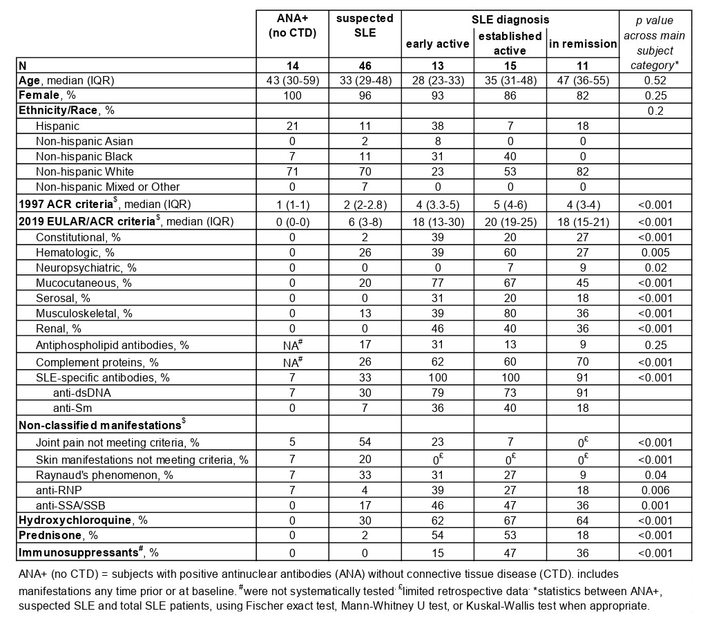
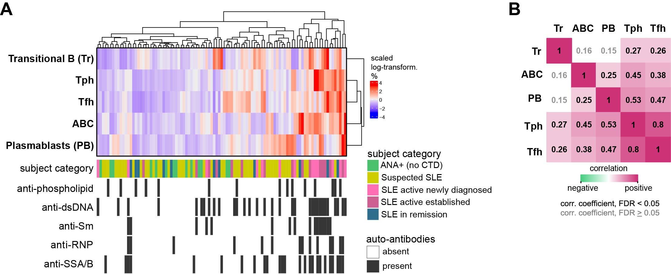
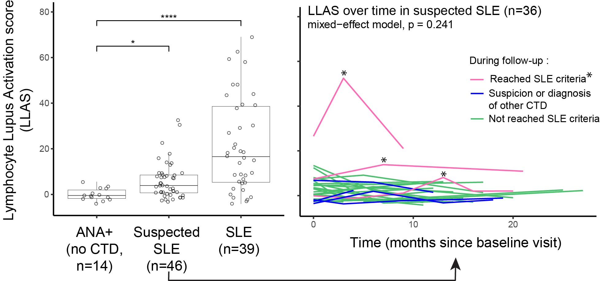
Results: We studied 145 samples: 103 from 46 patients with suspected SLE, 14 from ANA+ individuals, and 39 from patients with definite SLE (13 with active new SLE, 15 with active established disease, and 11 in remission) (Table 1 Baseline characteristics). 36 of the 46 patients with suspected SLE had multiple visits, mean (SD) follow-up 13.2 (5.6) months. In follow-up, 3 patients were diagnosed and met 2012 SLICC or 2019 EULAR/ACR criteria; 4 patients were diagnosed or suspected of having Sjögren’s syndrome, dermatomyositis, seronegative rheumatoid arthritis or spondylarthritis. To examine patterns of T/B cell proportions, we used hierarchical clustering and identified a gradient ranging from lowest proportions of all 5 cell subsets among ANA+ patients to the highest among active SLE patients with multiple autoantibodies (Figure 1A). Those with suspected SLE were widely distributed in the hierarchical clustering (Figure 1A). In the heatmap gradient and autoantibody-associated patterns, Tph and Tfh cells strongly correlated with ABCs and plasmablasts (Figure 1B). Patients with suspected SLE had significantly higher LLAS than ANA+ patients, yet with a broad range (Figure 2A). The 3 patients who developed classified SLE had either the highest or most increased LLAS in follow-up (Figure 2B).

Conclusion: Using flow cytometry with limited markers, we defined a LLAS score capturing key lymphocyte subsets implicated in SLE immune dysregulation. LLAS was highly expanded in SLE patients and in patients developing SLE (vs. ANA+ only patients), suggesting a role for LLAS as an early SLE biomarker.

Supporting image 1

Table 1. Baseline characteristics

Supporting image 2

Figure 1. Proportion of pathologic T-B cell phenotypes in ANA+ individuals, suspected and definite SLE patients. A. Heatmap of hierarchical clustering of proportions of transitional B cells (Tr), autoimmune-associated B cells (ABC), plasmablasts (PB), T peripheral helper (Tph) and T follicular helper (Tfh) in 99 baseline samples. Each column represents a subject’s sample. B. Spearman’s rho correlations between T and B cell subsets, including all 99 baseline samples.

Supporting image 3

Figure 2. Lymphocyte Lupus Activation Score (LLAS) in subjects category. Left panel represent baseline values and statistics are performed using Kruskal-Wallis test followed by corrrection for multiple comparison by Dunn’s test (* p < 0.05, **** p < 0.0001). Right panel represents LLAS measured at baseline and each follow-up visit in 36 patients with suspected SLE (total 103 sample-visits).


Disclosures: A. Horisberger: None; I. Adejorin: None; J. Caldropoli: None; E. Dillon: None; E. Oakes: None; K. Marks: None; T. Sasaki: None; K. Costenbader: Amgen, 2, 5, AstraZeneca, 5, Bain HealthSciences, 2, Bristol-Myers Squibb(BMS), 2, Cabaletta Bio, 2, 5, Exagen, 5, Gilead, 5, Glaxo Smith Kline, 2, 5, Merck, 5; D. Rao: Amgen, 6, AnaptysBio, 2, AstraZeneca, 1, Bristol-Myers Squibb, 2, 5, GlaxoSmithKline, 2, HiFiBio, 2, Janssen, 5, Merck, 5, Scipher Medicine, 2.

To cite this abstract in AMA style:

Horisberger A, Adejorin I, Caldropoli J, Dillon E, Oakes E, Marks K, Sasaki T, Costenbader K, Rao D. Lupus Lymphocyte Activation Score of Pathologic T and B Cell Phenotypes Increased in Patients at Risk of Systemic Lupus Erythematosus [abstract]. Arthritis Rheumatol. 2024; 76 (suppl 9). https://acrabstracts.org/abstract/lupus-lymphocyte-activation-score-of-pathologic-t-and-b-cell-phenotypes-increased-in-patients-at-risk-of-systemic-lupus-erythematosus/. Accessed .
  • Tweet
  • Click to email a link to a friend (Opens in new window) Email
  • Click to print (Opens in new window) Print

« Back to ACR Convergence 2024

ACR Meeting Abstracts - https://acrabstracts.org/abstract/lupus-lymphocyte-activation-score-of-pathologic-t-and-b-cell-phenotypes-increased-in-patients-at-risk-of-systemic-lupus-erythematosus/

Advanced Search

Your Favorites

You can save and print a list of your favorite abstracts during your browser session by clicking the “Favorite” button at the bottom of any abstract. View your favorites »

All abstracts accepted to ACR Convergence are under media embargo once the ACR has notified presenters of their abstract’s acceptance. They may be presented at other meetings or published as manuscripts after this time but should not be discussed in non-scholarly venues or outlets. The following embargo policies are strictly enforced by the ACR.

Accepted abstracts are made available to the public online in advance of the meeting and are published in a special online supplement of our scientific journal, Arthritis & Rheumatology. Information contained in those abstracts may not be released until the abstracts appear online. In an exception to the media embargo, academic institutions, private organizations, and companies with products whose value may be influenced by information contained in an abstract may issue a press release to coincide with the availability of an ACR abstract on the ACR website. However, the ACR continues to require that information that goes beyond that contained in the abstract (e.g., discussion of the abstract done as part of editorial news coverage) is under media embargo until 10:00 AM ET on November 14, 2024. Journalists with access to embargoed information cannot release articles or editorial news coverage before this time. Editorial news coverage is considered original articles/videos developed by employed journalists to report facts, commentary, and subject matter expert quotes in a narrative form using a variety of sources (e.g., research, announcements, press releases, events, etc.).

Violation of this policy may result in the abstract being withdrawn from the meeting and other measures deemed appropriate. Authors are responsible for notifying colleagues, institutions, communications firms, and all other stakeholders related to the development or promotion of the abstract about this policy. If you have questions about the ACR abstract embargo policy, please contact ACR abstracts staff at [email protected].

Wiley

  • Online Journal
  • Privacy Policy
  • Permissions Policies
  • Cookie Preferences

© Copyright 2025 American College of Rheumatology