ACR Meeting Abstracts

ACR Meeting Abstracts

  • Meetings
    • ACR Convergence 2024
    • ACR Convergence 2023
    • 2023 ACR/ARP PRSYM
    • ACR Convergence 2022
    • ACR Convergence 2021
    • ACR Convergence 2020
    • 2020 ACR/ARP PRSYM
    • 2019 ACR/ARP Annual Meeting
    • 2018-2009 Meetings
    • Download Abstracts
  • Keyword Index
  • Advanced Search
  • Your Favorites
    • Favorites
    • Login
    • View and print all favorites
    • Clear all your favorites
  • ACR Meetings

Abstract Number: 1250

Local Inflammatory Response Mediated by the Homing of Tfh17 Cells Is Involved in Tissue Injury of Immunoglobulin a Vasculitis

Xiaoxue Ma, Qinglian Jiang and Xuyang Chi, The First Hospital of China Medical University, Shenyang, China

Meeting: ACR Convergence 2023

Keywords: T Cell, Vasculitis

  • Tweet
  • Click to email a link to a friend (Opens in new window) Email
  • Click to print (Opens in new window) Print
Session Information

Date: Monday, November 13, 2023

Title: (1221–1255) Pediatric Rheumatology – Clinical Poster II: Connective Tissue Disease

Session Type: Poster Session B

Session Time: 9:00AM-11:00AM

Background/Purpose: Immunoglobulin A vasculitis (IgAV), also named Henoch–Schönlein purpura, is a systemic vasculitis characterized by the deposition of IgA1-dominant immune complexes in small vessels that often involves the skin, joints, gastrointestinal tract, and kidney. Research indicated increased frequencies of circulating activated B cells and plasmablasts in IgAV, which may serve as the source of
the rising IgA levels. T follicular helper (Tfh) 17 cells are considered to support B cells to switch to high-affinity IgA production. Previous study has confirmed that Tfh17 cells increase in the peripheral blood of IgAV patients. To evaluate the pathological role and the contribution in local tissue damage of Tfh17 cells in IgAV, we investigated the mechanism responsible for the differentiation of Tfh17 and the production of IgA in IgAV patients and IgAV rats respectively, and explored how to ameliorate IgAV by modulating Tfh17 generation.

Methods: Renal biopsy samples and peripheral blood mononuclear cells from IgAV patients were analyzed respectively by immunofluorescence staining and flow cytometry. In vitro culture was performed to assess the modulation of cytokine-induced phenotypes. IgAV rat model was established by intragastric administration of mixed solution, intraperitoneal injection of ovalbumin and Freund’s adjuvant. IgAV rats were used to explore the therapeutic effects of regulating Tfh17 cells. Serum cytokine and IgA levels were measured by ELISA while histopathological changes were evaluated by H&E and PAS staining. Flow cytometry and immunofluorescence staining were used to detect T cell and GC B cell phenotypes and homing characteristics in circulation and tissues of IgAV rats.

Results: Frequency of CD4+CXCR5+CCR6+Tfh17 cells were increased in the circulation and organization of IgAV patients and associated with disease severity. The increased expressions of CD103, CCR6, CCR9 lead to the homing and residence characteristics of Tfh17 cells, resulting in local inflammatory response. Suppression of Tfh17 cells reduced the production of IgA and greatly ameliorated clinical symptoms and decreased IgA deposition and mesangial proliferation in the kidney in IgAV rats.

Conclusion: Our findings suggest that suppression of Tfh17 differentiation can alleviate IgA-mediated vasculitis and inflammatory tissue damage, which may permit the development of tailored medicines for treating IgAV.

Supporting image 1

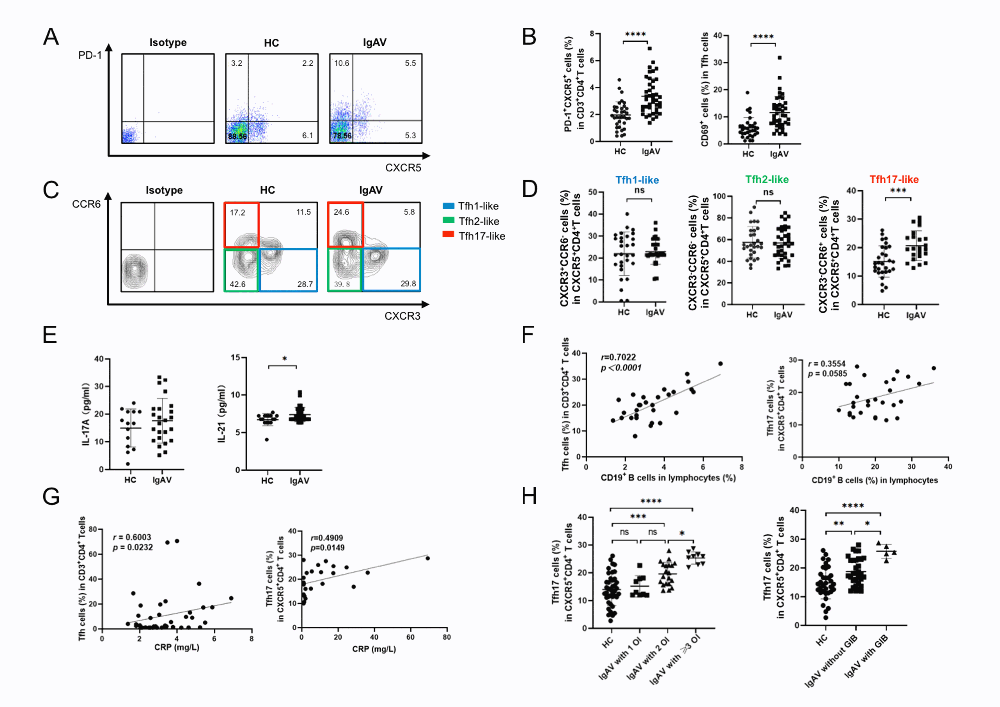
Circulating CD4+CXCR5+CCR6+ Tfh17 cells are increased and associated with IgAV disease severity. PBMCs and serum were isolated from the peripheral blood of IgAV patients and HCs without incubation. (A) Representative flow cytometry plots showing the percentage of circulating PD_1+CXCR5+ Tfh cells in CD3+CD4+ T cells. (B) Scatter plots showing the frequencies of circulating PD_1+CXCR5+ Tfh and CD69+ activated cTfh cells in CD3+CD4+ T cells. (C, D) Representative flow cytometry plots and scatter plots showing the percentages of cTfh1, cTfh2, and cTfh17 subsets in circulating CD3+CD4+CXCR5+ Tfh cells. (E) Serum levels of IL_17A and IL_21 evaluated by ELISA (25 of 40 IgAV patients were randomly selected and tested). Correlations of (F) frequencies of CD19+ B cells in lymphocytes (32 of 40 IgAV patients were randomly selected and tested) and (G) serum CRP levels with the percentages of cTfh cells and cTfh17 cells from IgAV patients. (H) Frequencies of circulating CXCR3−CCR6+ Tfh17 cells in circulating CD3+CD4+CXCR5+ Tfh cells from HC and IgAV patients grouped by the organ involvement (OI) number or the presence of gastrointestinal bleeding (GIB). ns: not significant; *p<0.05; **p<0.01; ***p<0.001; ****p<0.0001.

Supporting image 2

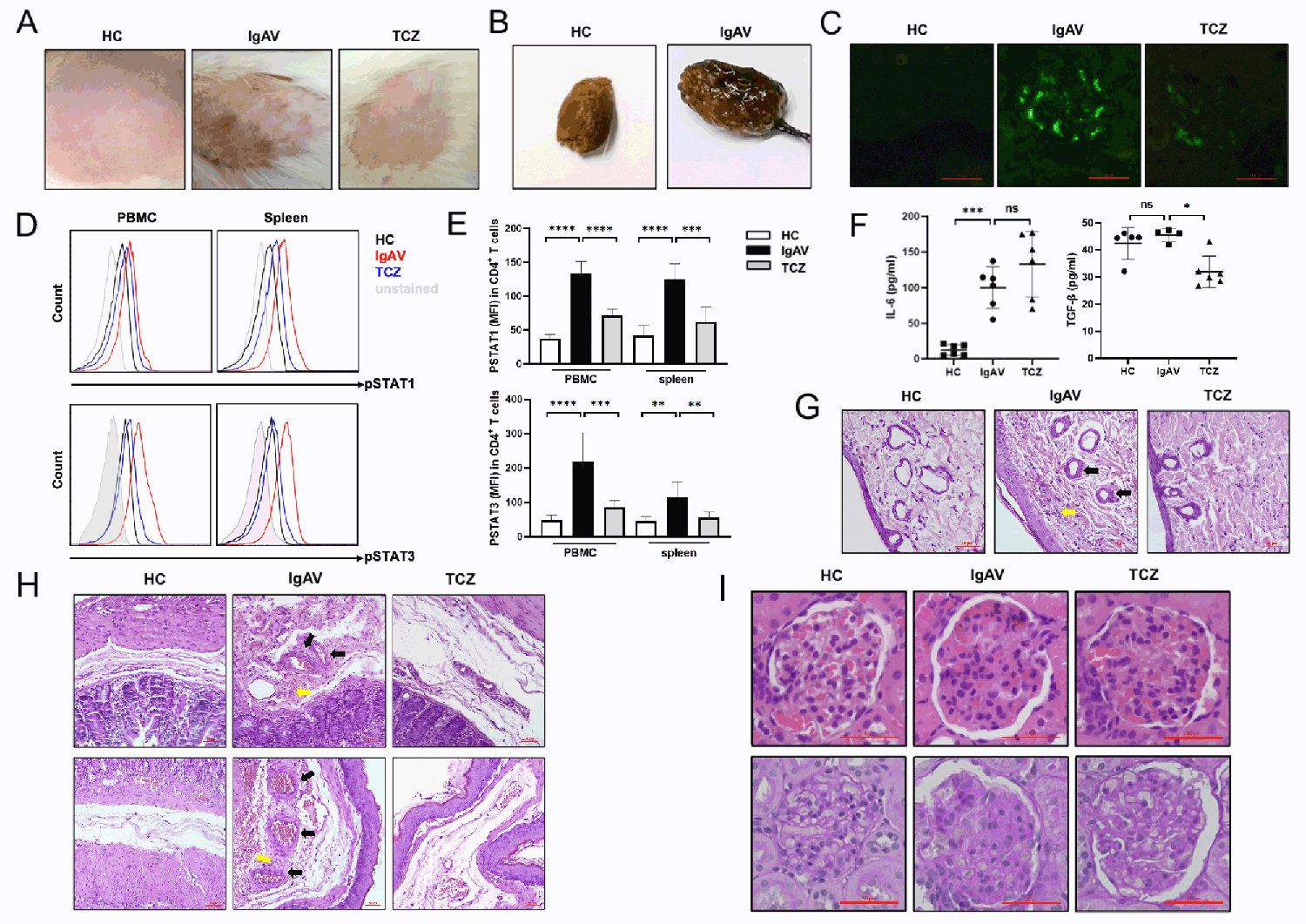
Suppressing Tfh17 using tocilizumab ameliorates symptoms in the IgAV rat. SD rats were respectively assigned to HC, IgAV, and TCZ groups. Representative photographs showing (A) hemorrhagic rash and (B) bloody stool from the three groups. Representative immunofluorescence images (C) showing renal IgA deposition from the three groups. (D, E) PBMCs and spleen cells were isolated from the three groups. Representative flow cytometry line graphs (D) and bar graphs (E) showing the expression of pSTAT1 and pSTAT3 in CD4+ T cells. (F) Serum levels of IL-6 and TGF-β in the three groups (the serum TGF-β level was tested in 5 rats from the HC group, 4 rats from the IgAV group, and 6 rats from the TCZ group). Representative photomicrographs showing H&E-stained sections of (G) skin and (H) intestinal (upper) and gastric (lower) tissues from the three groups. Black arrows indicate thickened vessel walls, hemangiectasis, and hyperemia; yellow arrows indicate extravasated erythrocytes. (I) Representative photomicrographs showing H&E-stained (upper) and PAS-stained (lower) sections of the kidney from the three groups. Scale bar = 50 μm. MFI: mean fluorescence intensity; ns: not significant; *p<0.05; **p<0.01; ***p<0.001; ****p<0.0001.

Supporting image 3

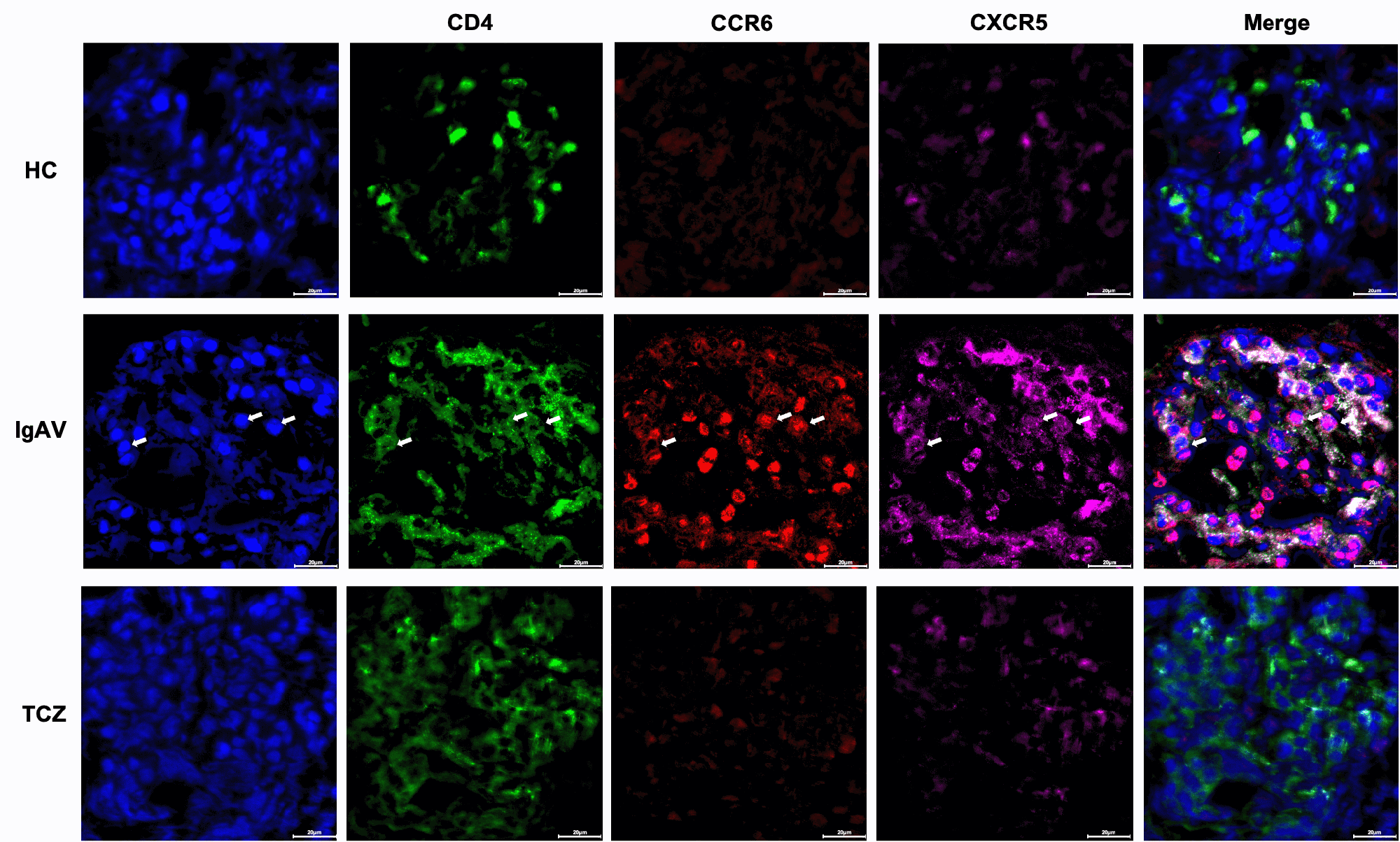
Expression of Tfh17 cells in the renal tissue of IgAV rat. Multichannel confocal
immunofluorescence staining of human renal tissue for CD4, CCR6 and CXCR5 with composite multiplexed images (scale bar: 20 lm). Arrowheads
indicate representative CD4+CCR6+ CXCR5+ Tfh17 cells.


Disclosures: X. Ma: None; Q. Jiang: None; X. Chi: None.

To cite this abstract in AMA style:

Ma X, Jiang Q, Chi X. Local Inflammatory Response Mediated by the Homing of Tfh17 Cells Is Involved in Tissue Injury of Immunoglobulin a Vasculitis [abstract]. Arthritis Rheumatol. 2023; 75 (suppl 9). https://acrabstracts.org/abstract/local-inflammatory-response-mediated-by-the-homing-of-tfh17-cells-is-involved-in-tissue-injury-of-immunoglobulin-a-vasculitis/. Accessed .
  • Tweet
  • Click to email a link to a friend (Opens in new window) Email
  • Click to print (Opens in new window) Print

« Back to ACR Convergence 2023

ACR Meeting Abstracts - https://acrabstracts.org/abstract/local-inflammatory-response-mediated-by-the-homing-of-tfh17-cells-is-involved-in-tissue-injury-of-immunoglobulin-a-vasculitis/

Advanced Search

Your Favorites

You can save and print a list of your favorite abstracts during your browser session by clicking the “Favorite” button at the bottom of any abstract. View your favorites »

All abstracts accepted to ACR Convergence are under media embargo once the ACR has notified presenters of their abstract’s acceptance. They may be presented at other meetings or published as manuscripts after this time but should not be discussed in non-scholarly venues or outlets. The following embargo policies are strictly enforced by the ACR.

Accepted abstracts are made available to the public online in advance of the meeting and are published in a special online supplement of our scientific journal, Arthritis & Rheumatology. Information contained in those abstracts may not be released until the abstracts appear online. In an exception to the media embargo, academic institutions, private organizations, and companies with products whose value may be influenced by information contained in an abstract may issue a press release to coincide with the availability of an ACR abstract on the ACR website. However, the ACR continues to require that information that goes beyond that contained in the abstract (e.g., discussion of the abstract done as part of editorial news coverage) is under media embargo until 10:00 AM ET on November 14, 2024. Journalists with access to embargoed information cannot release articles or editorial news coverage before this time. Editorial news coverage is considered original articles/videos developed by employed journalists to report facts, commentary, and subject matter expert quotes in a narrative form using a variety of sources (e.g., research, announcements, press releases, events, etc.).

Violation of this policy may result in the abstract being withdrawn from the meeting and other measures deemed appropriate. Authors are responsible for notifying colleagues, institutions, communications firms, and all other stakeholders related to the development or promotion of the abstract about this policy. If you have questions about the ACR abstract embargo policy, please contact ACR abstracts staff at [email protected].

Wiley

  • Online Journal
  • Privacy Policy
  • Permissions Policies
  • Cookie Preferences

© Copyright 2025 American College of Rheumatology