ACR Meeting Abstracts

ACR Meeting Abstracts

  • Meetings
    • ACR Convergence 2024
    • ACR Convergence 2023
    • 2023 ACR/ARP PRSYM
    • ACR Convergence 2022
    • ACR Convergence 2021
    • ACR Convergence 2020
    • 2020 ACR/ARP PRSYM
    • 2019 ACR/ARP Annual Meeting
    • 2018-2009 Meetings
    • Download Abstracts
  • Keyword Index
  • Advanced Search
  • Your Favorites
    • Favorites
    • Login
    • View and print all favorites
    • Clear all your favorites
  • ACR Meetings

Abstract Number: 1452

Inflammatory Markers and Left Ventricular Dysfunction in Systemic Lupus Erythematosus (SLE)

Audrey Hagiwara, Moumita Bose, Marianne Bernardo, Michael Nelson, Mariko Ishimori, Daniel Berman, C. Noel Bairey Merz, janet wei and Caroline Jefferies, Cedars-Sinai Medical Center, Los Angeles, CA

Meeting: ACR Convergence 2023

Keywords: Cardiovascular, Damage Index, Disease Activity, Systemic lupus erythematosus (SLE)

  • Tweet
  • Email
  • Print
Session Information

Date: Monday, November 13, 2023

Title: (1442–1487) SLE – Diagnosis, Manifestations, & Outcomes Poster II

Session Type: Poster Session B

Session Time: 9:00AM-11:00AM

Background/Purpose: Women with SLE have an elevated risk of cardiovascular disease. Many women with SLE frequently report chest pain in the absence of obstructive coronary artery disease (CAD) due to coronary microvascular dysfunction (CMD), a form of ischemia with no obstructive CAD (Manchanda et al, 2022). Echocardiographic studies have shown that SLE patients have reduced left ventricular (LV) function, which may also correlate with higher SLE disease activity scores (Gegenava et al, 2020). As such, we used cardiac magnetic resonance imaging (cMRI) to investigate the relationship between SLE, related inflammatory biomarkers, and cardiac function in female SLE patients.

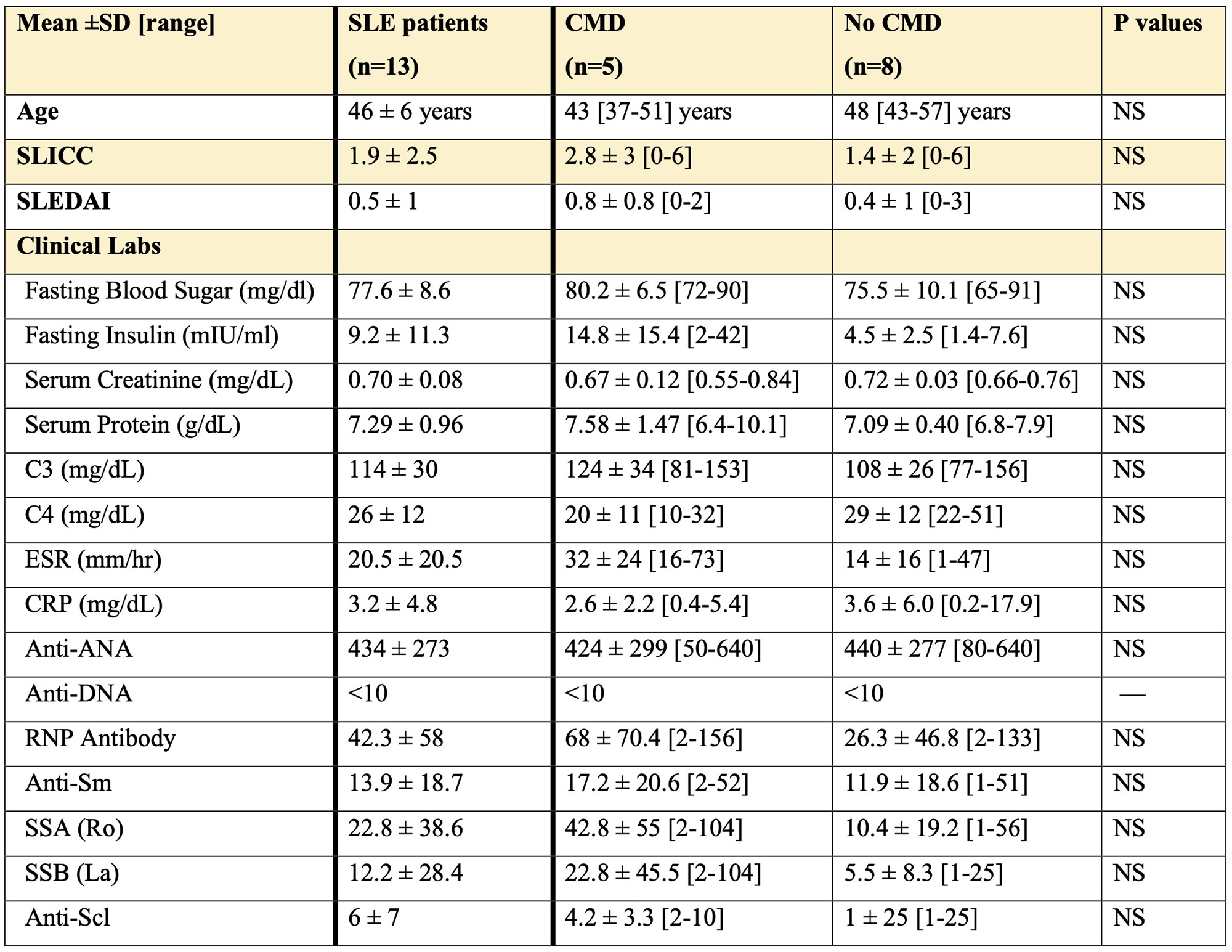
Methods: We performed stress cMRI in women with SLE and chest pain with no obstructive CAD (n=13, all met ACR 1997 criteria, Table 1) and reference controls (n=22) using our published protocol (Aldiwani et al, 2022). We evaluated LV function, tissue characterization (T1 mapping, ECV), and delayed enhancement, using CV142 software (Circle Cardiovascular Imaging Inc, Calgary, AB, Canada). Myocardial perfusion reserve index (MPRI) was calculated using our published protocol (Thomson et al, 2015). SLEDAI and SLICC Damage Index (DI) were calculated per validated criteria (Bombardier et al, 1992, Gladman et al, 1997). Serum samples were analyzed for inflammatory markers and autoantibodies (Table 1). Independent two-tailed t test was performed on clinical values with CMD and no CMD SLE subjects, and on cMRI values with all SLE subjects and controls. Correlation analysis was done on clinical values, and cMRI values on all SLE subjects.

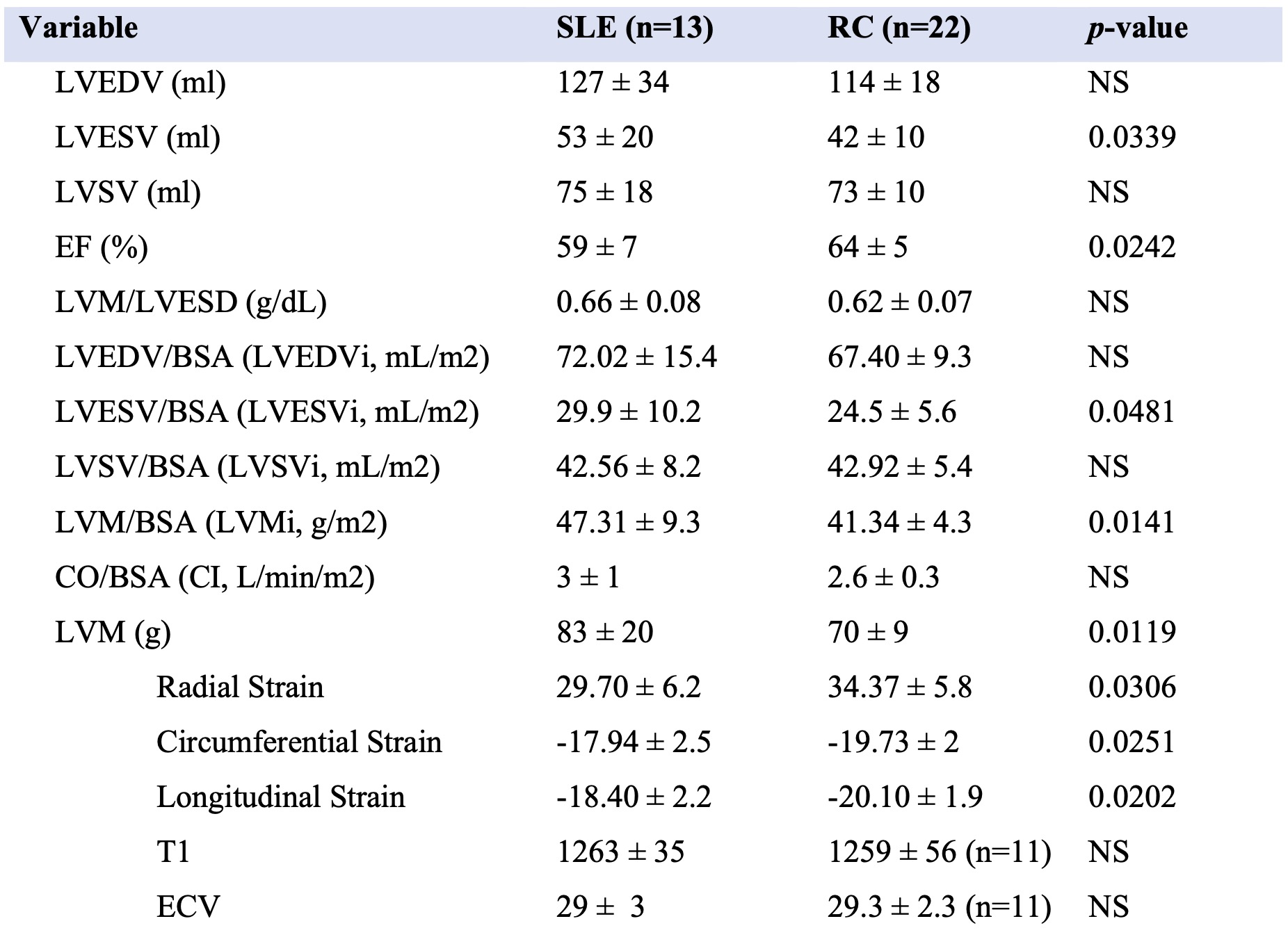
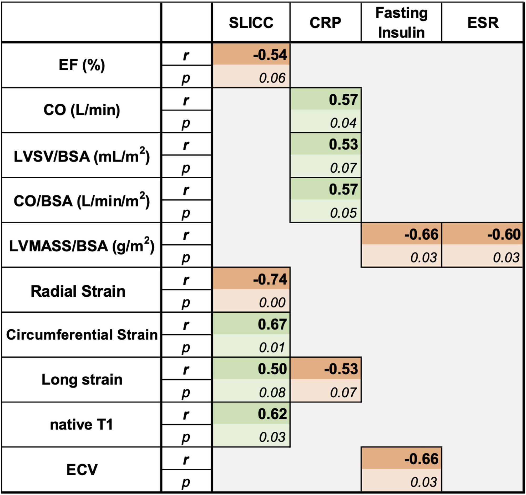
Results: Overall, 40% of SLE subjects had MPRI values < 1.84, consistent with CMD. Compared to controls, SLE subjects had significantly lower LVEF, and higher LVESVi and LVMi (Table 2). Corresponding to this, radial, longitudinal, and circumferential strain were significantly lower in the SLE subjects. In correlation analysis of serum inflammatory biomarkers to cMRI values in the SLE subjects, SLICC DI was related to worse cardiac function (lower radial, circumferential and longitudinal strain) and higher T1 time (Table 3). Additionally, fasting insulin and ESR were negatively correlated with LVMi. Fasting insulin also negatively correlated with ECV. CRP had a positive association with LVESV index and CI and a negative association with longitudinal strain.

Conclusion: Among women with SLE with chest pain and no obstructive CAD, 40% have CMD. While evaluations of known inflammatory markers (such as ESR and CRP) predictably correlated with decreased cardiac function (Lertratanakul et al, 2014), our study found that decreased fasting insulin levels is a novel marker associated with diminished LV function. In addition, although studies have used SLEDAI as a marker of disease activity when assessing cardiac dysfunction, we are the first to demonstrate that SLICC DI, an assessment of SLE damage, is also correlated with cardiac dysfunction in SLE. This indicates that SLE patients with higher SLICC DI and increased SLE-related damage could potentially have silent involvement in their cardiac tissue, and as such, SLICC DI is another tool that can be used to evaluate the association between SLE and LV dysfunction.

Supporting image 1

Table 1: Baseline Characteristics of Female SLE Participants. There was no group difference in clinical and laboratory values in the SLE participants with and without CMD. Complement 3 (C3); Complement 4 (C4); Erythrocyte Sedimentation Rate (ESR); C-Reactive Protein (CRP); Antinuclear antibody (ANA); Ribonucleoprotein (RNP); Smith (Sm); Anti-topoisomerase I antibody (Scl-70).

Supporting image 2

Table 2: Comparison of Cardiac MRI Analysis between SLE Participants and Reference Controls (RC). Left Ventricular (LV) End Diastolic Volume (LVEDV); LV End Systolic Volume (LVESV); LV Systolic Volume (LVSV); Ejection Fraction (EF); LV Mass (LVM); LV End Systolic Diameter (LVESD); Body Surface Area (BSA); LVEDV/BSA (LVEDV index); LVESV/BSA (LVESV index); LVSV/BSA (LVSV index); LVM/BSA (LVM index); CO/BSA (cardiac index), Extracellular Volume Fraction (ECV).

Supporting image 3

Table 3: Correlation analysis of cardiac function versus clinical values from SLE participants in the study. The Spearman r coefficient (r) and p value (p) is shown. Ejection Fraction (EF); Cardiac Output (CO); Left Ventricular (LV) Systolic Volume (LVSV); LV mass (LVM); Body Surface Area (BSA); Extracellular Volume (ECV); Systemic Lupus International Collaborating Clinics (SLICC) Damage Index; C-reactive Protein (CRP); Erythrocyte Sedimentation Rate (ESR).


Disclosures: A. Hagiwara: None; M. Bose: None; M. Bernardo: None; M. Nelson: None; M. Ishimori: None; D. Berman: None; C. Bairey Merz: None; j. wei: None; C. Jefferies: None.

To cite this abstract in AMA style:

Hagiwara A, Bose M, Bernardo M, Nelson M, Ishimori M, Berman D, Bairey Merz C, wei j, Jefferies C. Inflammatory Markers and Left Ventricular Dysfunction in Systemic Lupus Erythematosus (SLE) [abstract]. Arthritis Rheumatol. 2023; 75 (suppl 9). https://acrabstracts.org/abstract/inflammatory-markers-and-left-ventricular-dysfunction-in-systemic-lupus-erythematosus-sle/. Accessed .
  • Tweet
  • Email
  • Print

« Back to ACR Convergence 2023

ACR Meeting Abstracts - https://acrabstracts.org/abstract/inflammatory-markers-and-left-ventricular-dysfunction-in-systemic-lupus-erythematosus-sle/

Advanced Search

Your Favorites

You can save and print a list of your favorite abstracts during your browser session by clicking the “Favorite” button at the bottom of any abstract. View your favorites »

All abstracts accepted to ACR Convergence are under media embargo once the ACR has notified presenters of their abstract’s acceptance. They may be presented at other meetings or published as manuscripts after this time but should not be discussed in non-scholarly venues or outlets. The following embargo policies are strictly enforced by the ACR.

Accepted abstracts are made available to the public online in advance of the meeting and are published in a special online supplement of our scientific journal, Arthritis & Rheumatology. Information contained in those abstracts may not be released until the abstracts appear online. In an exception to the media embargo, academic institutions, private organizations, and companies with products whose value may be influenced by information contained in an abstract may issue a press release to coincide with the availability of an ACR abstract on the ACR website. However, the ACR continues to require that information that goes beyond that contained in the abstract (e.g., discussion of the abstract done as part of editorial news coverage) is under media embargo until 10:00 AM ET on November 14, 2024. Journalists with access to embargoed information cannot release articles or editorial news coverage before this time. Editorial news coverage is considered original articles/videos developed by employed journalists to report facts, commentary, and subject matter expert quotes in a narrative form using a variety of sources (e.g., research, announcements, press releases, events, etc.).

Violation of this policy may result in the abstract being withdrawn from the meeting and other measures deemed appropriate. Authors are responsible for notifying colleagues, institutions, communications firms, and all other stakeholders related to the development or promotion of the abstract about this policy. If you have questions about the ACR abstract embargo policy, please contact ACR abstracts staff at [email protected].

Wiley

  • Online Journal
  • Privacy Policy
  • Permissions Policies
  • Cookie Preferences

© Copyright 2025 American College of Rheumatology