Session Information
Session Type: Abstract Submissions (ACR)
Background/Purpose
Normal bones present cortical micro-channels (CMC), which carry micro-vessels and ensure communication between the bone marrow and the synovial compartment. Only recently these structures have been visualized in vivo in rheumatoid arthritis patients through high resolution peripheral computed tomography (HR-pQCT). We hypothesized that it is possible to detect and characterize these channels in healthy subjects (HS) and subjects with cutaneous psoriasis (PSO), assuming that PSO patients could present changes in bone microstructure previous to the onset of joint involvement.
Methods
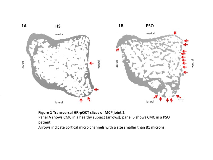
HR-pQCT (XtremeCT, Scanco Medical, Switzerland) scanning of the dominant hand was carried out on PSO patients without history of synovitis, dactylitis or enthesitis and HS. Image resolution was set at 82 μm voxel size. Images of the 2nd metacarpal head (MCH2) were downsized to the minimum of one slice with the 3D evaluation program provided by the manufacturer. Transversal, sagittal and coronal images were obtained using the subdim feature of the 3D evaluation program. Transversal slices (Fig.1) were projected at the level of the insertion of the joint capsule. Sagittal and coronal planes were set exactly into the middle of the MCH2. Demographic and clinical data were collected for the two groups of subjects. The study was conducted upon approval by the local ethic committee and the National Radiation Safety Agency (BfS). Patients participated after signing informed consent.
Results
PSO patients (N=56,M/F: 36/20, mean age 47.0±13.8 y) were compared to HS (N=24, M/F: 10/14, mean age 43.8±11.9 y). The subjects were comparable per age and sex. PSO patients exhibited moderately severe disease (PASI 7.9±8.9) and disease duration of 12.7±14.3 y, the most frequent phenotype being psoriasis vulgaris (78.6%), 60.7% presented scalp lesions, 48.2% had nail lesions. Imaging analysis was blindly performed by two readers. Inter- and intra-reader reliability was high (r=0.96 and r=0.98). In PSO patients 607 CMC were found vs. 97 in HS in the MCH2. Expressed as mean number of CMC this accounts for 10.8±9.1 vs. 4.1±3.7 (p<0.001). No correlation was found for the CMC number in the PSO patients and the severity of the cutaneous disease and its duration nor for the age of the subjects.
Conclusion
Visualization of cortical pathologies as small as 81 microns can be obtained by this technique. These images resemble rather histopathologic slices in vitro (Fig. 1). In our study cutaneous psoriatic patients with no clinical history of arthritis showed a significant higher number of these cortical channels compared to healthy subjects, suggesting an increased communication between bone marrow and joint in this phase of disease previous to the onset of joint involvement.
Disclosure:
D. Simon,
None;
F. Faustini,
None;
A. Kleyer,
None;
J. Haschka,
None;
D. Werner,
None;
A. J. Hueber,
None;
M. Sticherling,
None;
G. Schett,
None;
J. Rech,
None.
« Back to 2014 ACR/ARHP Annual Meeting
ACR Meeting Abstracts - https://acrabstracts.org/abstract/in-vivo-visualization-of-cortical-microchannels-in-metacarpal-bones-in-patients-with-cutaneous-psoriasis-by-high-resolution-peripheral-computed-tomography-detecting-cortical-pathologies-b/

