ACR Meeting Abstracts

ACR Meeting Abstracts

  • Meetings
    • ACR Convergence 2024
    • ACR Convergence 2023
    • 2023 ACR/ARP PRSYM
    • ACR Convergence 2022
    • ACR Convergence 2021
    • ACR Convergence 2020
    • 2020 ACR/ARP PRSYM
    • 2019 ACR/ARP Annual Meeting
    • 2018-2009 Meetings
    • Download Abstracts
  • Keyword Index
  • Advanced Search
  • Your Favorites
    • Favorites
    • Login
    • View and print all favorites
    • Clear all your favorites
  • ACR Meetings

Abstract Number: 1038

Improved Assessment of Structural Damage in the Spine in Patients with Axial Spondyloarthritis by MRI-based Synthetic CT: A Comparison with Low-dose CT and X-ray

Simone Tromborg Willesen1, Nora Vladimirova1, Jakob Moeller2, Anna Enevold Fløistrup Hadsbjerg1, Bimal Mayur Kumar Vora3, Sengul Seven1, Susanne Juhl Pedersen1 and Mikkel Østergaard1, 1Copenhagen Center for Arthritis Research, Center for Rheumatology and Spine Diseases, Centre for Head and Orthopaedics, Rigshospitalet; University of Copenhagen, Copenhagen, Denmark, 2Herlev-Gentofte Hospital, Herlev, Denmark, 3Singapore General Hospital, Singapore, Singapore

Meeting: ACR Convergence 2023

Keywords: Computed tomography (CT), Magnetic resonance imaging (MRI), Outcome measures, spondyloarthritis, X-ray

  • Tweet
  • Click to email a link to a friend (Opens in new window) Email
  • Click to print (Opens in new window) Print
Session Information

Date: Monday, November 13, 2023

Title: (1033–1051) Imaging of Rheumatic Diseases Poster I

Session Type: Poster Session B

Session Time: 9:00AM-11:00AM

Background/Purpose: Whether a therapy has bone structure-modifying effects in the spine is crucial to determine since this is a prerequisite for a therapy to reduce development of permanent disability in patients with axial spondyloarthritis (axSpA). The current imaging modality for assessing structural progression in axSpA is conventional radiography (X-ray), using the modified Stoke Ankylosing Spondylitis Structural Score (mSASSS). However, this method is only able to show change over a minimum of two years, i.e. has a very low sensitivity to change. Alternative modalities are MRI and CT. MRI, unfortunately, cannot reliably visualize small syndesmophytes. CT is considered the gold standard but delivers substantial ionizing radiation. However, modern CT scanners allow the use of lower radiation doses (low-dose CT, ldCT).

The purpose of this study was, with ldCT as the gold standard reference, to assess and compare the ability of a novel deep-learning MRI-based method (“synthetic CT”, sCT), ldCT, and X-ray, in the assessment of structural damage in the spine in patients with axSpA.

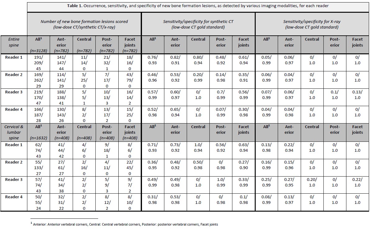
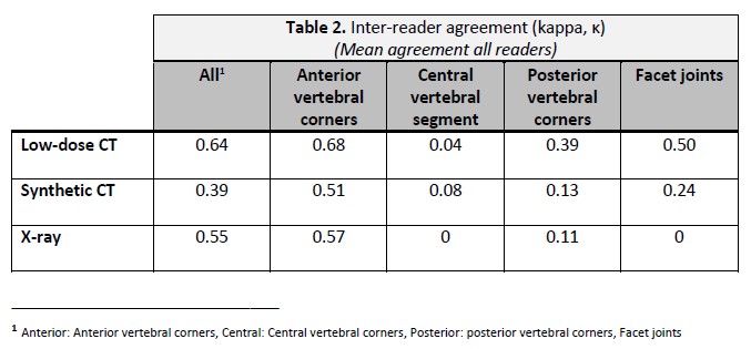
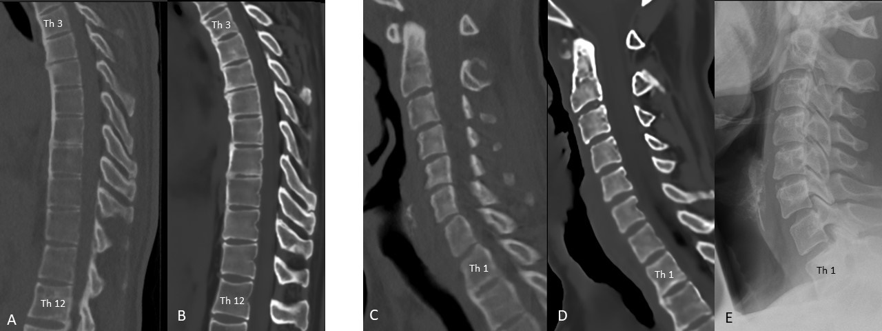
Methods: X-ray (lumbar and cervical spine), ldCT, and sCT of the entire spine were performed in patients with axSpA (according to ASAS criteria) in a prospective observational cohort. sCT was made using the BoneMRI application (v1.6, MRIGuidance BV, Utrecht, NL) a quantitative 3D MRI technique based on a multiple gradient-echo sequence and a machine learning processing pipeline that can generate CT-like MR images. All images were scored by four readers for new bone formation (NBF; such as syndesmophytes and ankylosis). NBF was recorded as present vs. absent at the anterior, central, and posterior locations as defined by the Canada Denmark (CANDEN) MRI scoring system for the spine and at the facet joints. Images were anonymized and read blinded to information from other imaging modalities and to clinical information. Readers were trained before the exercise by mutual evaluation of 3 other ldCT and sCT image sets. Sensitivity and specificity of sCT and X-ray were calculated, with ldCT as the gold standard. Inter-reader agreement was assessed using kappa statistics.

Results: 17 patients with axSpA were included (8 women, 9 men; mean age 34.4 ± 9.5 [SD]). The mean overall number of NBF lesions scored was 186 for ldCT, 207 for sCT, and 37 for X-ray. Most findings were located at the anterior vertebral corners with a mean of 143 lesions on ldCT, 142 on sCT, and 35 on X-ray (Table 1). With ldCT as the reference, the overall sensitivity for sCT to detect NBF was 0.58, with a corresponding specificity of 0.97. sCT had the highest sensitivity (0.65) at the anterior corners (specificity 0.94). For levels that were assessable on X-ray (C2-Th1 and Th12-S1), the sensitivity was 0.47 for sCT and 0.16 for X-ray, with similar high specificities ( >0.90) for both methods.

The mean inter-reader agreement for all locations was 0.64 for ldCT, 0.39 for sCT, and 0.55 for X-ray, and best for the anterior corners (Table 2).

Conclusion: MRI-based synthetic CT showed very high specificity and a sensitivity much higher than X-ray, despite limited reader training. With more training, calibration, and optimization of the scoring procedure, sCT could become highly valuable in axSpA clinical trials.

Supporting image 1

Supporting image 2

Supporting image 3

Figure 1: A-B: Low-dose CT (A) and MRI-based synthetic CT (sCT, B) of the thoracic spine. Ankylosis is seen anteriorly at several upper thoracic vertebral levels, with both modalities. C-E: Low-dose CT (C), sCT (D), and X-ray (E) of the cervical spine. Anterior ankylosis is seen at the Th1/Th2 level, both on low-dose CT and sCT, while this level is not visualized adequately by X-ray. Note: Low-dose CT versus sCT slice positioning (A-D) differ slightly at some levels. All images are from the same axSpA patient.


Disclosures: S. Tromborg Willesen: None; N. Vladimirova: None; J. Moeller: None; A. Hadsbjerg: None; B. Vora: None; S. Seven: None; S. Pedersen: Novartis, 5; M. Østergaard: AbbVie, 2, 5, 6, Amgen, 5, Boehringer-Ingelheim, 2, 6, Bristol-Myers Squibb(BMS), 2, 5, 6, Celgene, 2, 5, 6, Eli Lilly, 2, 6, Galapagos, 2, 6, Gilead, 2, 6, Hospira, 2, 6, Janssen, 2, 6, MEDAC, 6, Merck, 2, 5, 6, Novartis, 2, 5, 6, Novo Nordisk, 2, 6, Orion, 2, 6, Pfizer, 2, 6, Regeneron, 2, 6, Roche, 2, 6, Sandoz, 2, 6, Sanofi, 2, 6, UCB, 2, 6.

To cite this abstract in AMA style:

Tromborg Willesen S, Vladimirova N, Moeller J, Hadsbjerg A, Vora B, Seven S, Pedersen S, Østergaard M. Improved Assessment of Structural Damage in the Spine in Patients with Axial Spondyloarthritis by MRI-based Synthetic CT: A Comparison with Low-dose CT and X-ray [abstract]. Arthritis Rheumatol. 2023; 75 (suppl 9). https://acrabstracts.org/abstract/improved-assessment-of-structural-damage-in-the-spine-in-patients-with-axial-spondyloarthritis-by-mri-based-synthetic-ct-a-comparison-with-low-dose-ct-and-x-ray/. Accessed .
  • Tweet
  • Click to email a link to a friend (Opens in new window) Email
  • Click to print (Opens in new window) Print

« Back to ACR Convergence 2023

ACR Meeting Abstracts - https://acrabstracts.org/abstract/improved-assessment-of-structural-damage-in-the-spine-in-patients-with-axial-spondyloarthritis-by-mri-based-synthetic-ct-a-comparison-with-low-dose-ct-and-x-ray/

Advanced Search

Your Favorites

You can save and print a list of your favorite abstracts during your browser session by clicking the “Favorite” button at the bottom of any abstract. View your favorites »

All abstracts accepted to ACR Convergence are under media embargo once the ACR has notified presenters of their abstract’s acceptance. They may be presented at other meetings or published as manuscripts after this time but should not be discussed in non-scholarly venues or outlets. The following embargo policies are strictly enforced by the ACR.

Accepted abstracts are made available to the public online in advance of the meeting and are published in a special online supplement of our scientific journal, Arthritis & Rheumatology. Information contained in those abstracts may not be released until the abstracts appear online. In an exception to the media embargo, academic institutions, private organizations, and companies with products whose value may be influenced by information contained in an abstract may issue a press release to coincide with the availability of an ACR abstract on the ACR website. However, the ACR continues to require that information that goes beyond that contained in the abstract (e.g., discussion of the abstract done as part of editorial news coverage) is under media embargo until 10:00 AM ET on November 14, 2024. Journalists with access to embargoed information cannot release articles or editorial news coverage before this time. Editorial news coverage is considered original articles/videos developed by employed journalists to report facts, commentary, and subject matter expert quotes in a narrative form using a variety of sources (e.g., research, announcements, press releases, events, etc.).

Violation of this policy may result in the abstract being withdrawn from the meeting and other measures deemed appropriate. Authors are responsible for notifying colleagues, institutions, communications firms, and all other stakeholders related to the development or promotion of the abstract about this policy. If you have questions about the ACR abstract embargo policy, please contact ACR abstracts staff at [email protected].

Wiley

  • Online Journal
  • Privacy Policy
  • Permissions Policies
  • Cookie Preferences

© Copyright 2025 American College of Rheumatology