Background/Purpose: Precise quantification of joint space morphology (JSM) is an important factor for early diagnosis and monitoring of disease activity and therapeutic responses in rheumatoid arthritis (RA). Patients with active RA usually present painful, stiff and swollen joints and have difficulties in stretching their joints. Positioning of these patients is a major concern for accurate assessment as it is responsible for an important variability in cross-sectional and longitudinal studies.
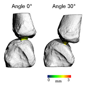
We have developed a method to characterize 3D JSM of the metacarpophalangeal (MCP) joint by high resolution peripheral quantitative computed tomography (HR-pQCT). We assessed the impact on JSM of measurements performed with a 0° and a 30° flexion of the MCP joints, which we assume would be convenient for most active RA patients.
Methods: We performed HR-pQCT on the second metacarpophalangeal (MCP) joints of twelve ex vivo hands (3 men, 3 women, mean age = 77±9 yrs) and developed an automated algorithm to compute joint space volume (JSV), mean, minimal and maximal joint space width (JSW, JSW.MIN, JSW.MAX), as well as its distribution (JSW.SD) and asymmetry (JSW.AS, ratio of JSW.MAX over JSW.MIN). MCPs were scanned with a 0° and a 30° degree angle, with an inclined custom cast. The effects of different hand positioning were assessed by paired t-test.
Results: Angles between metacarpal and phalangeal bones were retrospectively verified on the reconstructed 3D images, with mean values of 5.2±4.5° (ranging from 0° to 12°) and 29.5±3.7° (ranging from 25° to 35°).
Positioning markedly impacted the mean and maximum values of joint space width. JSW was 16.4% thicker (2.00±0.38 vs. 1.72±0.30 mm; p=0.001) and JSW.MAX was increased by 12.5% (2.32±0.34 vs. 2.07±0.34 mm; p=0.001) when measured at 30° compared to 0° respectively. Its impact was less marked on joint space volume (64±22 vs. 59±17 mm3; 8.9%; p=0.29), minimum joint space width (1.28±0.22 vs. 1.18±0.31 mm; 8.7%; p=0.23) and asymmetry of joint space width (1.85±0.30 vs. 1.68±0.42; 10.2%; p=0.09), at 30° compared to 0° respectively. The heterogeneity of the joint space width (JSW.SD) was not affected by hand positions (0.22±0.09 vs. 0.21±0.08 mm; p=0.88).
Conclusion: This study suggests that positioning remains a major concern even with a 3D quantification of JSM. On the basis of active RA patient comfort, the use of a fixed-inclined cast would probably be the most appropriate method to accurately follow-up disease activity.
Disclosure:
S. Boutroy,
None;
E. Hirschenhahn,
None;
E. Youssof,
None;
H. Locrelle,
None;
T. Thomas,
None;
R. Chapurlat,
None;
H. Marotte,
None.
« Back to 2013 ACR/ARHP Annual Meeting
ACR Meeting Abstracts - https://acrabstracts.org/abstract/importance-of-hand-positioning-in-3d-joint-space-morphology-assessment/

