Session Information
Date: Monday, October 27, 2025
Title: (1553–1591) Systemic Sclerosis & Related Disorders – Clinical Poster II
Session Type: Poster Session B
Session Time: 10:30AM-12:30PM
Background/Purpose: Pulmonary fibrosis is a recognized sequela of severe COVID-19 pneumonia, but its additive impact on pre-existing interstitial lung disease (ILD)—particularly in patients with connective tissue disease (CTD)—remains poorly understood. This study aimed to assess the effect of prior COVID-19 infection on fibrosis progression in CTD-associated ILD (CTD-ILD) using artificial intelligence (AI)-based quantitative CT (QCT) analysis.
Methods: We conducted a single-center retrospective study of CTD-ILD patients with confirmed COVID-19 pneumonia who underwent at least two high-resolution CT (HRCT) scans: one within 18 months before infection and one ≥6 months after infection. Only follow-up scans obtained >1 year post-infection were included in the final analysis. Controls were CTD-ILD patients without a history of COVID-19, matched for baseline HRCT and follow-up interval (12–18 months). Fibrosis progression was quantified using both visual and AI-based QCT analysis (Figure 1). Clinical characteristics, pulmonary function tests (PFTs), and immunosuppressive treatment history were collected. Multivariable linear regression identified independent predictors of fibrosis progression.
Results: A total of 110 patients were included (COVID-19 group: n=53; control group: n=57). The cohort comprised various CTD subtypes: systemic sclerosis (54.7%), Sjögren’s syndrome (23.2%), rheumatoid arthritis (15.8%), polymyositis (3.2%), dermatomyositis (1.1%), and mixed connective tissue disease (2.1%). Baseline CTD subtype, COVID-19 treatment history, baseline PFTs, and initial fibrosis extent and pattern were comparable between groups. At follow-up, the COVID-19 group showed a significantly greater extent of reticulation on QCT (3.1% vs. 1.4%, p=0.044) and a trend toward increased honeycombing. Multivariable analysis revealed that prior COVID-19 infection (β=4.332, p=0.044), older age (β=0.211, p=0.004), lower baseline FVC (β=−0.187, p=0.001), and rituximab use (β=6.607, p=0.022) were independently associated with greater fibrosis scores (Table 1).
Conclusion: In CTD-ILD patients, prior COVID-19 infection was independently associated with increased fibrosis progression on follow-up CT. These findings highlight the long-term pulmonary impact of COVID-19 in a vulnerable population, warranting close radiologic surveillance and personalized management strategies.
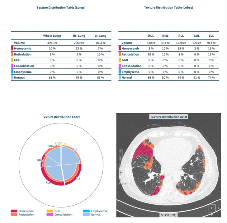 Figure 1. AI-based Quantitative CT (QCT) Analysis Example
Representative output of automated lung texture classification in a CTD-ILD patient using QCT. Distribution of honeycombing, reticulation, and normal parenchyma are quantified in tables (upper panels), a polar chart (lower left), and an axial HRCT image with overlaid classification (lower right). Color coding corresponds to specific radiologic textures (legend below chart).
.jpg)
Table 1. Linear Regression Analyses for Factors Associated with Increased Fibrosis Scores on AI-based QCT
To cite this abstract in AMA style:
Lee k, Kim H, Nam B. Impact of COVID-19 Infection on Fibrosis Progression in CTD-Associated ILD: An AI-Based Quantitative CT Study [abstract]. Arthritis Rheumatol. 2025; 77 (suppl 9). https://acrabstracts.org/abstract/impact-of-covid-19-infection-on-fibrosis-progression-in-ctd-associated-ild-an-ai-based-quantitative-ct-study/. Accessed .« Back to ACR Convergence 2025
ACR Meeting Abstracts - https://acrabstracts.org/abstract/impact-of-covid-19-infection-on-fibrosis-progression-in-ctd-associated-ild-an-ai-based-quantitative-ct-study/