Session Information
Session Type: Poster Session B
Session Time: 10:30AM-12:30PM
Background/Purpose: Cognitive impairment (CI) in systemic lupus erythematosus (SLE) may result from a chronic pro-inflammatory state in which immunosenescent and exhausted T-lymphocytes could be involved. This study investigates the expression of senescent and exhausted T cells markers in SLE patients with CI.
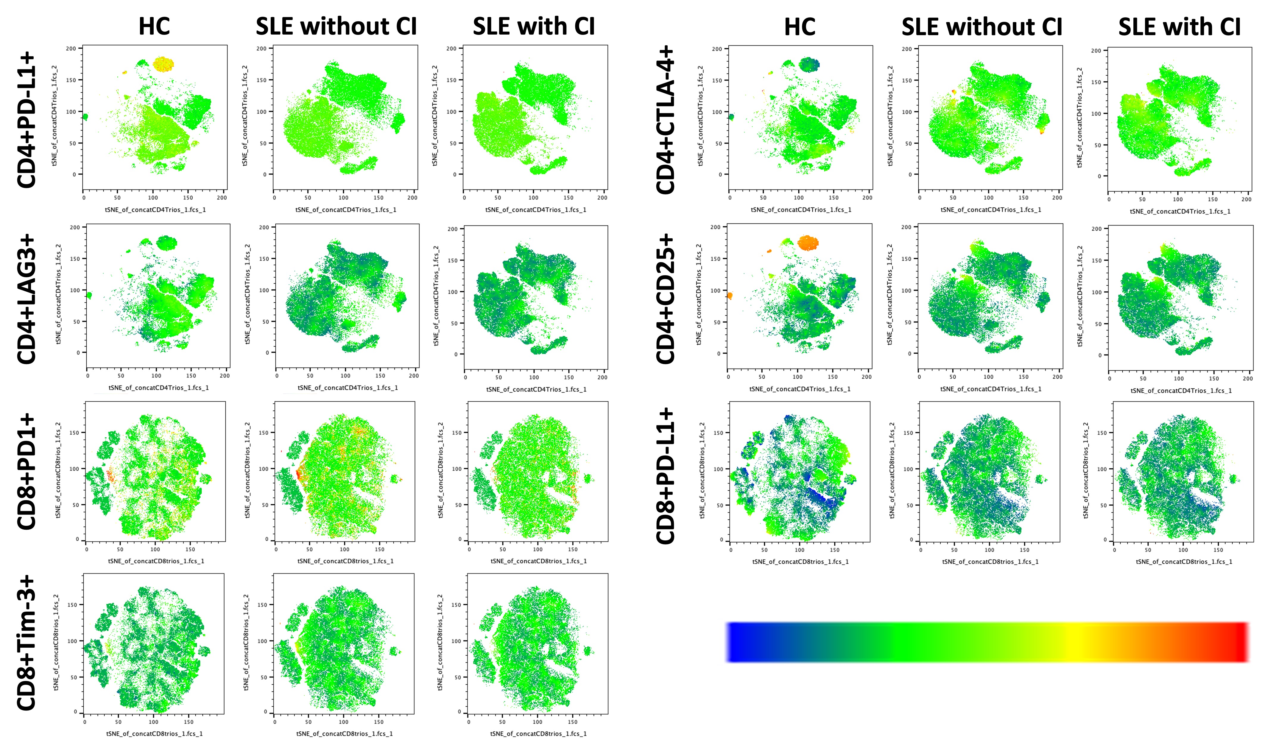
Methods: We included women aged 18-50 years, classified with SLE according to the EULAR/ACR 2019 criteria. We excluded women with other autoimmune diseases except antiphospholipid syndrome or Sjögren’s syndrome, administration of any biologic drug < 6 months prior to study entry, clinical SLEDAI-2K >0, SDI ≥1, current prednisone >7.5 mg/day, education level ≤6 years, pregnancy or premature ovarian failure, active infection, malignant disease, history of other neuropsychiatric manifestations, other chronic comorbidities or major depressive disorder. Healthy women (HC) paired for age ±5 years were also included. All patients underwent a neurocognitive battery applied by an expert neuropsychologist, assessing eight cognitive domains: concentration, verbal memory, visuospatial memory, language, processing speed, motor speed, problem solving and executive function. CI was defined when a subject had scores ≥2 standard deviation below the mean of the normative data in ≥1 cognitive domain. After the neurocognitive assessment patients were classified as: SLE with CI, SLE without CI and HC. Relative expression of senescence markers (CD27, CD28, CD57, KLRG1) and inhibitory markers (PD-1, PD-L1, Tim-3, CTLA-4 and LAG-3) in CD4+ and CD8+ lymphocytes were determined by flow cytometry, dimensionality reduction and clustering analysis. We generated tSNE maps using concatenated files with an equal number of CD3+ cells from eight representative trios of patients from each study group. Demographic and clinical data were recorded.
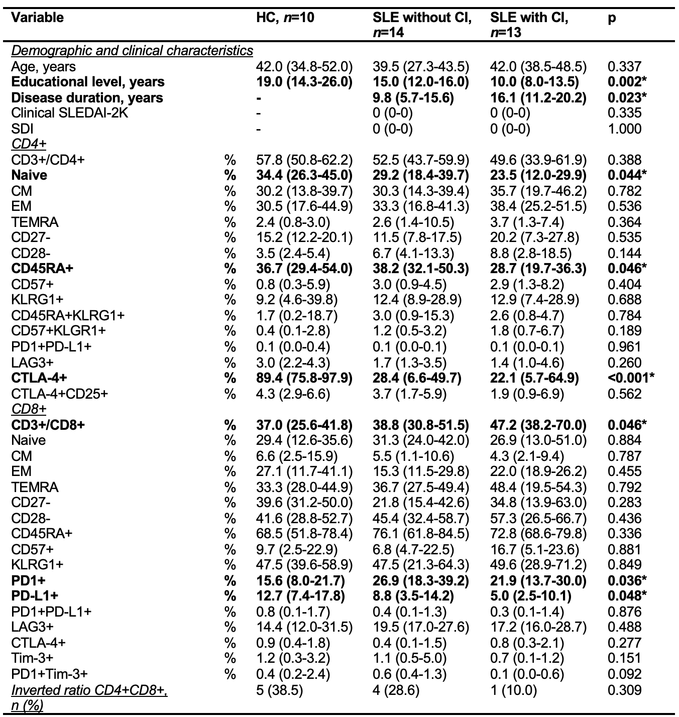
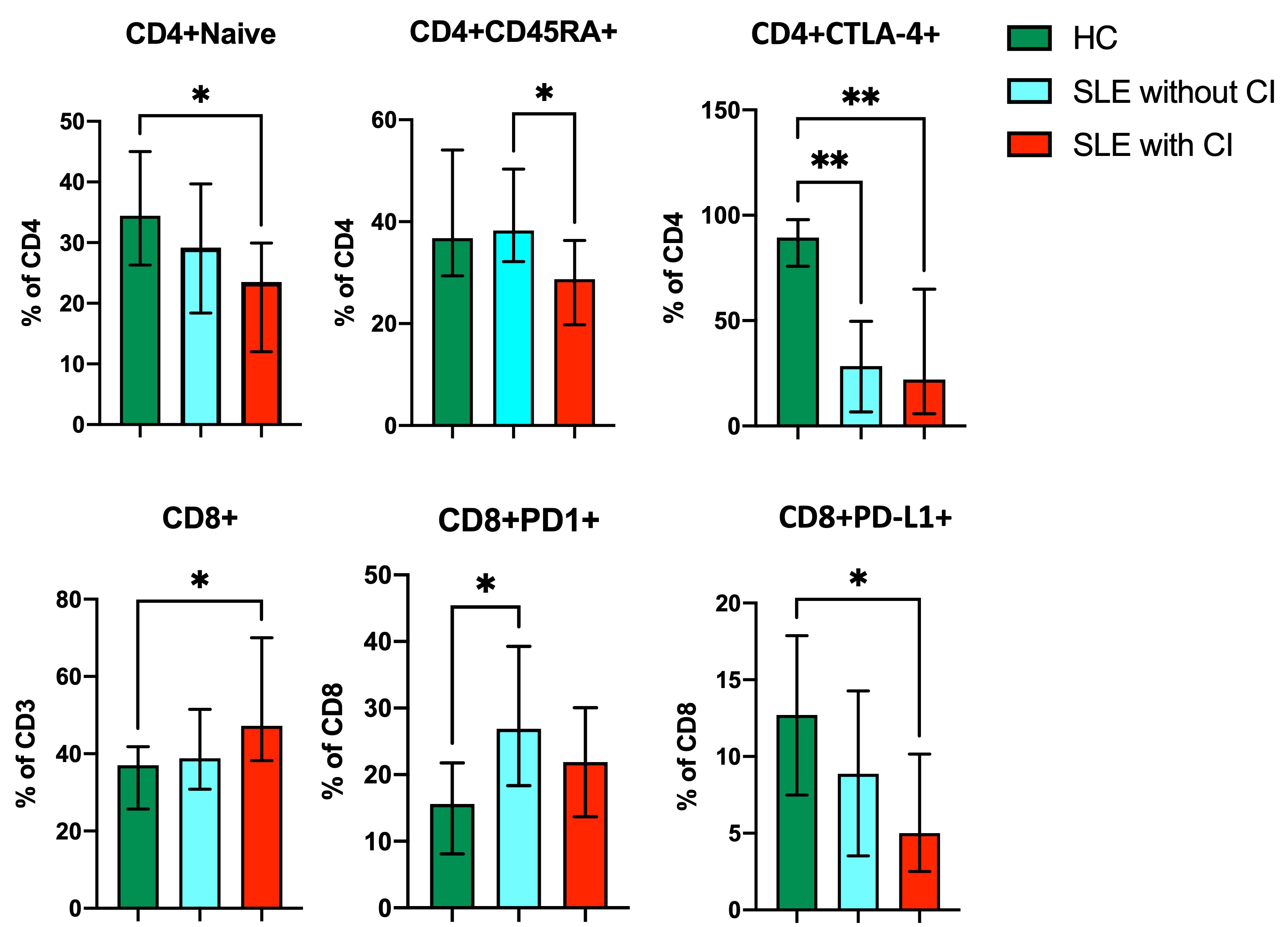
Results: Thirteen patients with SLE with CI, fourteen SLE without CI and ten HC were analyzed. The median age of patients studied was similar. SLE patients with CI had lower educational level than the SLE without CI and HC, and longer disease duration compared to SLE patients without CI. Disease activity and damage accrual was similar between the SLE groups (Table 1). In SLE patients with CI, the most frequently impaired cognitive domain was motor speed (76.9%), followed by visuospatial memory (30.8%), verbal memory (7.7%), language (7.7%), processing speed (7.7%) and problem solving (7.7%). SLE patients with CI had lower levels of CD4+ Naive, CD4+CD45RA+, CD4+CTLA-4+ and CD8+PD-L1+, but higher levels of CD8+ (Table 1, Figure 1). We could identify three representative CD8+ and four CD4+ subpopulations according to their relative expression of surface markers (Figure 2).
Conclusion: Significant differences were identified among the three groups in relation to markers of cellular exhaustion but not of immunosenescence. In SLE with CI, PD-L1, Tim-3, CTLA-4 and LAG-3 in CD4+ and Tim-3 and PD-L1 in CD8+ expression was lower compared to SLE without CI and HC. The study suggests the possibility that cellular exhaustion play a role in CI in SLE and could be useful for its diagnosis.
Data are expressed as proportions (percentages) or medians (interquartile ranges). Statistical analysis: Chi-square or Fisher’s exact tests were used for nominal variables, the Wilcoxon rank-sum test was used for numerical variables.
CM: central memory, EM: effector memory, TEMRA: effector memory T cells re-expressing CD45RA.
*Statistically significant.
HC: healthy control, SLE: systemic lupus erythematosus, CI: cognitive impairment.
HC: healthy control, SLE: systemic lupus erythematosus, CI: cognitive impairment.
To cite this abstract in AMA style:
Cimé-Aké E, Lima G, G. Lazarini E, Juárez S, Llorente L, Fragoso-Loyo H. Immunosenescent and Exhausted T Cells in Patients with Systemic Lupus Erythematosus and Cognitive Impairment [abstract]. Arthritis Rheumatol. 2024; 76 (suppl 9). https://acrabstracts.org/abstract/immunosenescent-and-exhausted-t-cells-in-patients-with-systemic-lupus-erythematosus-and-cognitive-impairment/. Accessed .« Back to ACR Convergence 2024
ACR Meeting Abstracts - https://acrabstracts.org/abstract/immunosenescent-and-exhausted-t-cells-in-patients-with-systemic-lupus-erythematosus-and-cognitive-impairment/


