Session Information
Session Type: Poster Session B
Session Time: 9:00AM-11:00AM
Background/Purpose: Synovial fluid analysis using polarized microscopy is the gold standard for the diagnosis of crystal-related arthritis. In our experience, we have noted that, when calcium pyrophosphate(CPP) crystals are observed, they sometimes appear within intracellular vacuoles. However, this phenomenon is not seen in those samples containing monosodium urate (MSU) crystals.This finding has been scantly reported in the literature, but may be useful in clinical practice to ensure accurate crystal identification.
Objectives: our study aims to assess hether the presence of vacuoles contributes to identifying the type of crystal, and also to gauge the frequency of their presentation.
Methods: We conducted an observational study in a rheumatology unit between February and June of 2019. Synovial fluids containing CPP or MSU crystals, obtained in daily clinical practice, were consecutively included for analysis. Two observers simultaneously analyzed the presence of vacuoles by ordinary light and phase contrast microscopy in less than 24 hours after their extraction, using a microscope equipped with two viewing stations. The primary study variable was to determine whether CPP and MSU crystals are seen inside intracellular vacuoles, and to calculate the frequency of this finding for each type of crystal, stimating their 95% confidence interval (95% CI) and comparing rates using Fisher’s exact test.
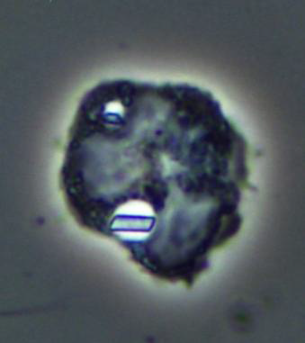
Results: Twenty-one samples were obtained. Data is given in the Table. MSU crystals were present in 7 (33.3%) and CPP crystals in 14 (66.6%). Interestingly, none of the MSU samples showed crystal-containing vacuoles (95% CI 0-35.4%). On the contrary, cytoplasmic vacuoles containing crystals were present in all of the CPP samples (95% CI 78.5-100%). The findings were confirmed by phase-contrast microscopy. Differences were statistically significant (p< 0.001).
Conclusion: The presence of vacuoles may be a useful and easy way to differentiate MSU and CPP crystals when performing synovial fluid microscopy in clinical practice, since it appears to be a distinctive feature in CPP crystal fluids.
 Image 1. Microscopy with ordinary light. Cells with cytoplasmic vacuoles are observed, as well as abundant intra and extracellular CPP crystals.
 Image 2. Microscopy with phase contrast technique. Cells with intracellular vacuoles are observed inside which have microcrystals with parallelepiped morphology, compatible with CPP.
To cite this abstract in AMA style:
Peral M, Calabuig I, Martín-Carratalá A, Andrés M, Pascual E. Identification of Intracellular Vacuoles in Synovial Fluid with Calcium Pyrophosphate and Monosodium Urate Crystals [abstract]. Arthritis Rheumatol. 2020; 72 (suppl 10). https://acrabstracts.org/abstract/identification-of-intracellular-vacuoles-in-synovial-fluid-with-calcium-pyrophosphate-and-monosodium-urate-crystals/. Accessed .« Back to ACR Convergence 2020
ACR Meeting Abstracts - https://acrabstracts.org/abstract/identification-of-intracellular-vacuoles-in-synovial-fluid-with-calcium-pyrophosphate-and-monosodium-urate-crystals/

