Session Information
Session Type: Poster Session C
Session Time: 1:00PM-3:00PM
Background/Purpose: Computer analysis of radiological images has been recently introduced in the study of patients with musculoskeletal pathologies. Our research group has shown that this type of ultrasound image analysis is sensitive to exercise-induced adaptive changes.
In the field of ultrasound, baseline settings and differences between images obtained by different equipment pose a potential problem in the comparative study of patients. The purpose of this study is to determine if the computer analysis of entheses ultrasound images can distinguish adaptive changes from those produced by spondyloarthritis.
Methods: A test validation study was carried out. Since the analysis of static ultrasound images strongly depends on the characteristics of the image and the ultrasound settings, three indices based on the findings in pathological territories and their counterpart in healthy entheses were defined.
Gray Intensity Mean Index (iMIG): Mean gray intensity in the clinically affected enthesis/mean gray intensity in the same healthy enthesis.
Gray Intensity Dispersion Index (iDIG): Mean of the standard deviation of the gray intensity means in the clinically affected enthesis/mean of the standard deviation of the gray intensity means in the same healthy enthesis.
Index of Gray Intensity Modes (iMoG): Mean gray intensity modes in the affected enthesis/mean gray intensity in the same healthy enthesis.
The same indices were used in the controls with the dominant side as the numerator and the non-dominant side as the denominator.
All the studied entheses corresponded to the lower limbs.
Results: Records of 49 patients (20 diagnosed with psoriatic arthritis, 19 with radiographic axial spondyloarthritis and 10 with non-radiographic axial spondyloarthritis) and 100 healthy controls were included. Of the 49 studies, 30 corresponded to the Achilles tendon, 10 to the distal patellar tendon and 9 to the proximal patellar tendon.
In the patient group, iMIG was 0.917 SD 0.062, iDIG was 0.976 SD 0.042, and iMoIG was 0.962 SD 0.041. In the control group, iMIG 1 SD 0.020, iDIG was 1.001 SD 0.03, and iMoIG was 0.999 SD 0.03.
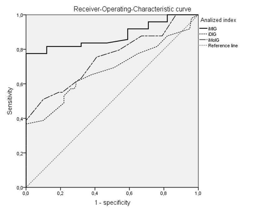
The area under the curve (figure 1) for the three indices was: iMIG 0.879 (95% CI 0.807 to 0.951), iDIG (95% CI 0.582 to 0.785), iMoIG 0.751 (95% CI 0.662 to 0.840). All areas demonstrated diagnostic validity with P< .0001.
According to the matrix of coordinates of the receiver-operating-characteristic (ROC) curve, for an iMIG cut-off point less than or equal to 1.02, a sensitivity of 95.9% and a specificity of 71% would be achieved.
Conclusion: The indices that contrast the means, dispersion and mode of gray intensities are valid for discriminating between healthy subjects and patients with load-bearing enthesopathies related to spondyloarthritis. Of all of them, the iMIG seems the most reliable. A cut-off point of less than 1.02 is proposed to identify patients with inflammatory enthesopathies.
In patients, the affected entheses have a lower mean gray than the healthy counterpart, so iMIG tends to be less than 1. In healthy subjects, the mean gray is slightly higher in the dominant enthesis, so iMIG tends to be greater than or equal to 1.
To cite this abstract in AMA style:
Tortosa Cabanas M, GUILLEN ASTETE C, Andreu Suarez A. Gray Intensity Ratio Indices as a Method of Discrimination Between Changes Induced by Sport and by Spondyloarthritis in Load-bearing Entheses [abstract]. Arthritis Rheumatol. 2022; 74 (suppl 9). https://acrabstracts.org/abstract/gray-intensity-ratio-indices-as-a-method-of-discrimination-between-changes-induced-by-sport-and-by-spondyloarthritis-in-load-bearing-entheses/. Accessed .« Back to ACR Convergence 2022
ACR Meeting Abstracts - https://acrabstracts.org/abstract/gray-intensity-ratio-indices-as-a-method-of-discrimination-between-changes-induced-by-sport-and-by-spondyloarthritis-in-load-bearing-entheses/

